
Adipocyte lipolysis research sits at a genuinely complex intersection of hormone signaling, lipid droplet biology, and metabolic disease modeling. Yet many labs running in vitro fat oxidation experiments are still working with incomplete pictures of the glucagon pathway, treating glucagon as a secondary or even irrelevant variable when studying triglyceride breakdown in white adipose cells. That is a significant oversight. The glucagon pathway does not just regulate hepatic glucose output. It directly engages the cAMP-PKA cascade inside adipocytes, controls access to lipid droplets through perilipin phosphorylation, and sits upstream of both ATGL and HSL activation. If your fat oxidation research model does not account for glucagon receptor inputs, your lipolysis readouts are incomplete by design.
Table of Contents
- Quick Takeaways
- The Lipolytic Cascade Inside Adipocytes
- How Glucagon Engages the Lipolytic Pathway
- Fat Oxidation Research Model Design
- Measuring Lipolysis In Vitro: What Actually Works
- Triple-Receptor Agonists and the New Complexity of Glucagon Pathway Research
- Comparison of In Vitro Adipocyte Lipolysis Research Approaches
- Compound Quality and Its Effect on Data Reliability
- Frequently Asked Questions
- References
Quick Takeaways
| Key Insight | Explanation |
|---|---|
| Glucagon activates lipolysis via cAMP/PKA | Glucagon receptor stimulation raises intracellular cAMP, activating PKA, which phosphorylates HSL and perilipins to initiate triglyceride hydrolysis. |
| ATGL requires CGI-58 dissociation from perilipin | PKA-mediated phosphorylation of perilipin causes CGI-58 to dissociate and activate ATGL, the primary lipase converting triglycerides to diacylglycerols. |
| Glucagon receptor expression in adipocytes is low but functional | Adipocytes express glucagon receptor mRNA and protein at low levels, making receptor density a critical experimental variable when modeling direct glucagon effects on fat cells. |
| Both glycerol and free fatty acid release must be measured | Measuring only one readout is insufficient. FFA re-uptake can distort results; glycerol is the more reliable sole indicator of net lipolysis when BSA buffering is suboptimal. |
| Hepatic vs. adipose glucagon signaling produce different outcomes | Recent research indicates that glucagon-induced energy expenditure operates primarily through hepatic receptor signaling, not adipose receptor signaling, a distinction critical for compartment-specific study design. |
| Triple agonists complicate single-pathway attribution | Compounds engaging GLP-1, GIP, and glucagon receptors simultaneously produce adipose tissue remodeling that cannot be attributed to any single receptor arm without careful controls. |
| Peptide purity is not optional in lipolysis assays | Impurities at the 1-5% level in research peptides can activate or suppress off-target receptors, producing cAMP artifacts that mimic or suppress glucagon-mediated lipolytic signals. |
The Lipolytic Cascade Inside Adipocytes
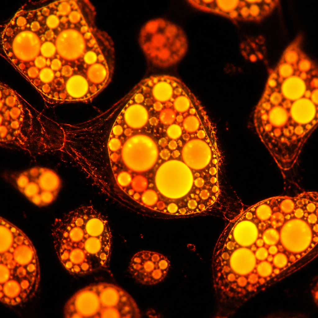
Adipocytes store energy as triglycerides in lipid droplets, and the architecture of those droplets is not passive. The lipid core is coated with perilipins, structural proteins whose primary job is to restrict access by lipases. Lipolysis does not begin until those guards are moved out of position.
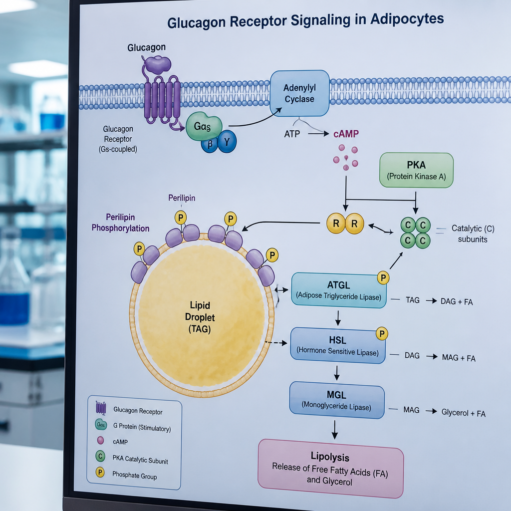
The signal that moves them is cAMP. When adenylate cyclase (AC) in the adipocyte plasma membrane is activated, intracellular cAMP rises and stimulates protein kinase A (PKA). PKA then phosphorylates two targets simultaneously: hormone-sensitive lipase (HSL) and the perilipins themselves. Phosphorylated perilipin releases CGI-58, which binds to and activates adipose triglyceride lipase (ATGL). ATGL converts triglycerides to diacylglycerols; HSL then converts those diacylglycerols to monoglycerols, with downstream lipases completing the hydrolysis to free glycerol and free fatty acids.
Circulating levels of free fatty acids (FFAs) and glycerol in a cell culture medium or a blood sample directly reflect the rate of this process. That is why both are used as primary readouts in adipocyte lipolysis research.
Pro tip: The cAMP-to-PKA axis is where most experimental manipulations either succeed or break down. If your PKA activation is inconsistent between replicates, check your adenylate cyclase stimulator concentration before re-examining receptor-level hypotheses.
How Glucagon Engages the Lipolytic Pathway
Glucagon's role in adipocyte biology is frequently undersold in experimental design, which is a practical problem. In the fasting state, glucagon secretion rises as insulin concentrations fall below the threshold needed to suppress lipolysis. At that point, glucagon and catecholamines converge on the same adenylate cyclase mechanism in fat cells, raising cAMP and triggering the PKA cascade described above.
The Direct Receptor Question
For glucagon to act directly on adipocytes, its receptor must be expressed on the cell surface. Glucagon receptor mRNA has been detected in rat adipocytes, and the debate in the field has centered on whether the receptor density is physiologically sufficient to mediate a meaningful direct lipolytic effect, or whether glucagon's effect on adipose lipolysis is mostly indirect, mediated through catecholamine release or hepatic signaling. Recent research provides important nuance here: glucagon-induced energy expenditure appears to operate primarily through hepatic glucagon receptor signaling rather than through adipose tissue receptors. This does not eliminate the adipocyte as a research target. It reframes what you are measuring when you stimulate adipocytes directly with a glucagon pathway peptide.
cAMP as the Central Variable
Whether glucagon acts directly or indirectly on the adipocyte, the cAMP concentration inside the cell is the functional variable that drives lipase activation. Insulin counteracts this by activating phosphodiesterase 3B (PDE3B), which degrades cAMP and suppresses lipolysis. Understanding this push-pull between cAMP-elevating signals (glucagon, catecholamines) and cAMP-degrading signals (insulin via PDE3B) is essential for interpreting any glucagon pathway experiment in fat cells. Recent data also implicates PDE4B and PDE4D downregulation as a mechanism by which sustained glucagon signaling maintains elevated cAMP even under conditions where receptor desensitization would otherwise occur.
In adipocyte lipolysis research, cAMP is not just a second messenger. It is the rate-controlling variable whose concentration determines the entire downstream lipase cascade. Failing to measure or control cAMP levels while modulating glucagon receptor activity produces uninterpretable data.
Pro tip: When designing glucagon pathway experiments, run a parallel cAMP ELISA on the same cells at each time point. This confirms that your peptide concentration is achieving receptor-level engagement and distinguishes receptor-mediated from non-specific lipolytic effects.
Fat Oxidation Research Model Design
Choosing the right fat oxidation research model for glucagon pathway work is not simply a matter of picking a cell line. The model choice determines what you can and cannot claim about the mechanism.
Primary Adipocytes vs. Cell Lines
Primary adipocytes differentiated from preadipocytes are highly lipolytic and maintain a more physiologically relevant receptor profile than established cell lines. 3T3-L1 adipocytes, the most widely used cell line, have notably lower basal lipolytic activity than primary cells and may underrepresent glucagon receptor density. If you are specifically studying glucagon receptor-mediated lipolysis, primary murine or human preadipocytes differentiated in vitro will give you more interpretable data than 3T3-L1 cells at baseline. 3T3-L1 is more appropriate for studying the downstream PKA-lipase machinery once the receptor engagement question has been addressed separately.
Ex Vivo Adipose Tissue as a Bridge Model
Ex vivo murine adipose tissue fragments preserve the three-dimensional tissue architecture, cell-cell contacts, and stromal vascular fraction that are absent in monolayer adipocyte cultures. For glucagon pathway work, this matters because paracrine signals from immune cells and preadipocytes within the tissue affect lipolytic tone. Ex vivo preparations are especially useful when testing glucagon pathway peptides that are known to have broad systemic effects, since the tissue context provides a more realistic response profile than isolated adipocytes. The trade-off is reduced experimental control over receptor expression and signal timing.
Key Controls Every Protocol Needs
Every well-designed adipocyte lipolysis experiment involving glucagon pathway modulation needs at minimum: a vehicle control, a maximal stimulation control (typically isoproterenol at saturating concentration), and an insulin suppression control. Without these three anchors, you cannot place your glucagon peptide response on the actual dynamic range of the system, and you will not know whether a blunted response reflects receptor antagonism, signal saturation, or simply poor cell viability.
Measuring Lipolysis In Vitro: What Actually Works
The practical execution of a lipolysis assay is where adipocyte lipolysis research most commonly falls apart. The biology is understood; the bench work is where inconsistency enters.
Glycerol vs. Free Fatty Acids: Which to Measure
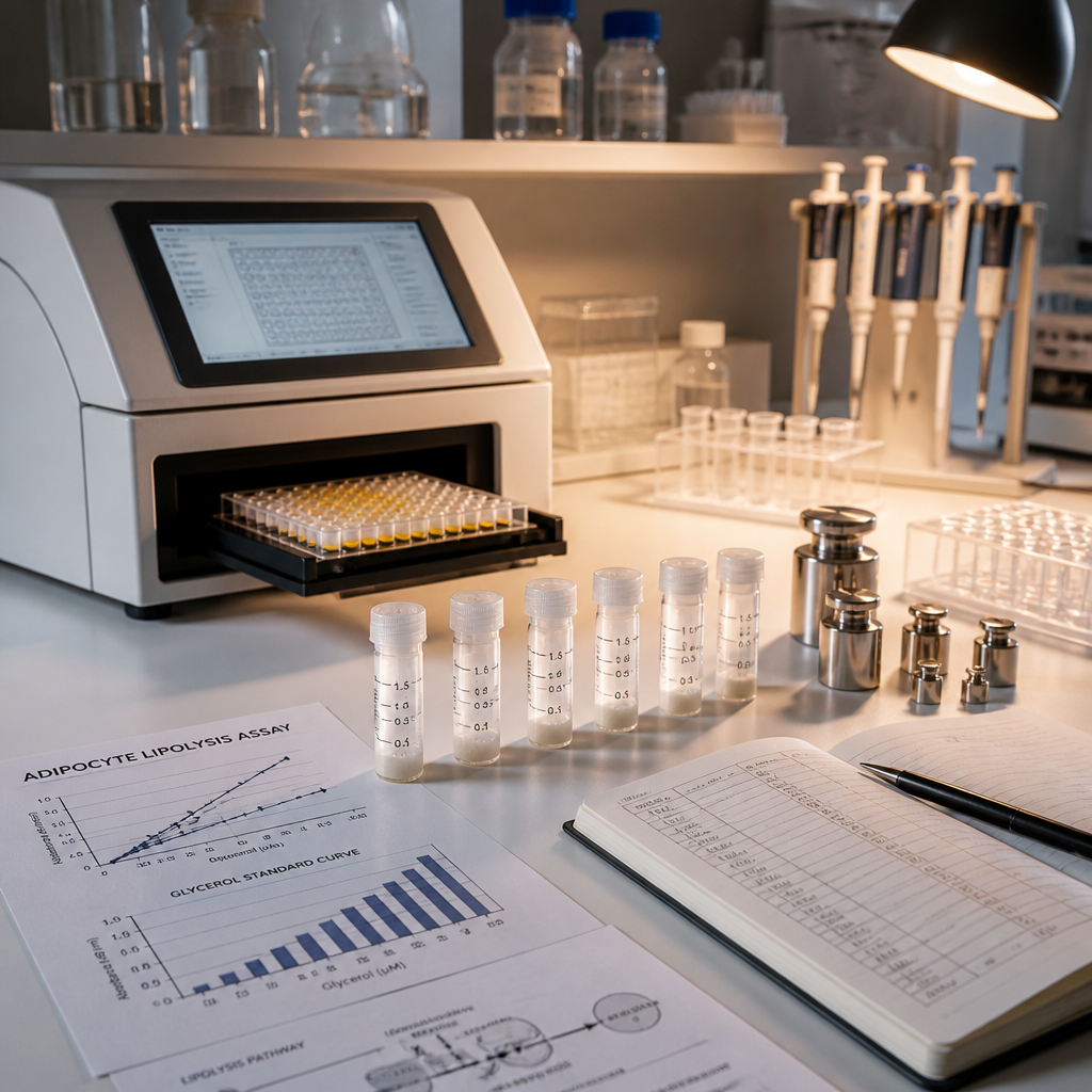
Both glycerol and FFA release into the culture medium are valid lipolysis readouts, and the ideal approach measures both at multiple time points to calculate a linear rate. Because white adipocytes express very little glycerol kinase, released glycerol is not re-taken up by the cell, making it a clean, accumulation-style readout. FFAs, by contrast, are subject to re-uptake depending on the albumin (BSA) concentration in the medium. If the FFA:glycerol ratio falls below 2:1 in your assay, you are losing FFAs back into cells or to insufficient BSA binding, and the FFA measurement is unreliable. The standard correction is to increase BSA concentration or collection frequency, not to re-run the entire experiment.
Time Point Selection and Rate Linearity
A common mistake in glucagon pathway lipolysis experiments is measuring a single end-point rather than a rate. Lipolysis is a dynamic process and the release of glycerol and FFAs follows a rate that can become non-linear as the assay extends beyond the linear phase. Collecting samples at 30-minute intervals and confirming linearity before reporting a rate value is not excessive caution. It is the difference between a publishable quantitative result and a directional finding that reviewers will correctly challenge.
Pro tip: Normalize all lipolysis rate data to total cellular protein or total DNA content from the same well. Variation in adipocyte differentiation efficiency between wells is common enough that unnormalized glycerol or FFA values will produce highly variable results even when your treatment effect is real and consistent.
Triple-Receptor Agonists and the New Complexity of Glucagon Pathway Research
The emergence of multi-receptor agonist peptides has substantially changed how glucagon pathway modulation is studied in adipose tissue. A glucagon receptor agonist operating alone produces a predictable cAMP-driven lipolytic response. A compound that simultaneously engages GLP-1, GIP, and glucagon receptors produces a layered response where the three receptor arms interact, and attributing any observed effect to the glucagon component specifically requires more sophisticated experimental design.
Research on retatrutide, a triple agonist of GIP, GLP-1, and glucagon receptors, has demonstrated multi-omic reprogramming of epididymal white adipose tissue, characterized by suppressed lipogenesis, enhanced fatty acid oxidation and mitochondrial function, restored peroxisomal activity, and downregulated inflammatory and fibrotic pathways. This is not a simple lipolysis-on/off result. It reflects a coordinated remodeling of adipose tissue metabolism that involves transcriptomic and metabolomic changes well beyond what a single-receptor model would predict.
For labs designing glucagon pathway peptide experiments that involve compounds with multi-receptor activity, this creates an important methodological requirement: receptor-selective controls. Running your multi-agonist alongside a glucagon receptor-selective agonist and a GLP-1 receptor-selective agonist as separate treatment arms allows component attribution. Without this, multi-omic results are descriptive rather than mechanistic.
The Glucagon Component in Adipose Remodeling
Glucagon functions beyond simple lipolysis stimulation. Its metabolic effects in the adipose compartment include reduced lipogenesis, maintenance of intracellular lipolysis through cAMP and PKA activity, and increased fatty acid oxidation. The glucagon receptor arm of triple agonists is generally considered to be the primary driver of elevated energy expenditure, particularly through hepatic pathways, distinguishing these compounds from GLP-1 mono-agonists that reduce energy intake without the same degree of thermogenic effect.
For research scientists at institutions studying metabolic disease, obesity biology, or MASLD, high-purity retatrutide peptide verified by Canadian laboratories and backed by full chain of custody documentation represents the most reliable starting point for this class of multi-receptor adipose tissue experiments.
Comparison of In Vitro Adipocyte Lipolysis Research Approaches
No single model covers all experimental questions in adipocyte lipolysis research. The three most commonly used approaches each have clearly defined strengths and failure modes when applied to glucagon pathway work specifically.
| Approach | Best Use Case for Glucagon Pathway Work | Key Limitation |
|---|---|---|
| 3T3-L1 Differentiated Adipocytes | Studying downstream PKA-ATGL-HSL cascade mechanics; high reproducibility across labs | Low basal lipolytic activity and potentially reduced glucagon receptor density; poor model for direct glucagon receptor-mediated lipolysis |
| Primary Preadipocytes Differentiated In Vitro | Receptor-level glucagon pathway work; physiologically relevant lipolytic rates; species-specific receptor expression studies | Higher batch-to-batch variability; requires optimization of differentiation efficiency; limited scale-up potential |
| Ex Vivo Adipose Tissue Fragments | Testing multi-receptor agonist peptides where tissue architecture and paracrine signaling influence the response; bridging in vitro to in vivo | Reduced experimental control over receptor expression; inter-animal variability; unsuitable for high-throughput compound screening |
Compound Quality and Its Effect on Data Reliability
This section exists because it addresses a problem that is not theoretical. Glucagon pathway peptides used in adipocyte lipolysis research are biologically active at nanomolar concentrations. At that sensitivity, a peptide preparation with 3-5% impurities is not a minor inconvenience. Truncated sequences, oxidized methionine residues, and aggregated peptide fractions can independently activate or suppress cAMP-coupled receptors, produce false-positive lipolysis signals, or suppress the response you are trying to measure. The data produced using such preparations does not reflect biology. It reflects chemistry.
Research-grade suppliers who provide independent laboratory verification, batch traceability, and documented chain of custody remove this ambiguity. When a lipolysis result is inconsistent between replicates from different peptide lots, the first diagnostic question is whether both lots have the same independently verified purity documentation. If the answer is no, the experiment has a confound that cannot be controlled through better statistical analysis.
For Canadian research institutions, sourcing peptides with Xpresspost-grade cold-chain delivery and lyophilized formulations further protects compound integrity between the supplier and the bench. Lyophilized peptides stored correctly before reconstitution avoid the degradation that liquid formulations can suffer in transit. This is especially relevant for glucagon pathway peptides, which contain structured motifs that are sensitive to hydrolysis and aggregation at non-optimal storage temperatures.
At Pepura Labs, every lyophilized peptide batch is independently verified by Canadian laboratories, with full batch traceability and chain of custody documentation provided to the researcher. For glucagon pathway lipolysis experiments where data integrity depends directly on compound purity, this verification is not a premium feature. It is the baseline standard that research reproducibility requires.
Frequently Asked Questions
Does glucagon directly stimulate lipolysis in human adipocytes?
The direct lipolytic effect of glucagon on adipocytes remains an area of active research. Glucagon receptor mRNA has been detected in adipocytes, and the signaling machinery (cAMP/PKA/ATGL/HSL) through which it would act is well established. However, recent research demonstrates that glucagon-induced increases in energy expenditure operate primarily through hepatic receptor signaling rather than adipose receptor signaling. Direct adipocyte lipolysis from glucagon receptor activation is likely but depends on receptor density, which varies by species, fat depot, and metabolic state. Experimental designs should test both direct and indirect (catecholamine-mediated) mechanisms.
What is the best readout for glucagon pathway lipolysis experiments in vitro?
Measuring both glycerol and free fatty acid release at multiple time points is the most informative approach. Glycerol is the preferred primary readout because it is not re-taken up by white adipocytes, giving a clean accumulation signal. FFAs provide a secondary confirmation but require sufficient BSA in the medium to prevent re-uptake. Confirm a linear release rate before reporting a rate value, and always normalize to total protein or DNA content from the same well.
Why is the FFA to glycerol ratio important in lipolysis assays?
Complete hydrolysis of one triglyceride molecule releases three fatty acids and one glycerol. A FFA:glycerol ratio below approximately 2:1 in your assay medium indicates that fatty acids are being re-taken up by cells or are bound insufficiently by BSA, meaning your FFA measurement is underestimating actual lipolysis. When this occurs, increasing BSA concentration, reducing collection intervals to every 30 minutes, or shortening the total assay duration to 2.5 hours are the standard corrections.
How should I control for the GLP-1 and GIP components when studying glucagon receptor effects in a multi-agonist experiment?
Include receptor-selective agonists as separate treatment arms alongside your multi-agonist compound. A glucagon receptor-selective agonist, a GLP-1 receptor-selective agonist, and a GIP receptor-selective agonist run in parallel at equimolar concentrations allow you to partition the observed effect. Receptor knockout cell models or selective antagonist co-treatments are a more stringent approach when the research question specifically requires component attribution rather than whole-compound characterization.
What cell model is most appropriate for studying glucagon pathway modulation of adipocyte lipolysis?
Primary preadipocytes differentiated in vitro are the most appropriate starting point for glucagon receptor-level work because they maintain a more physiologically relevant receptor profile and higher basal lipolytic rates than 3T3-L1 cells. 3T3-L1 cells are well suited to downstream PKA-lipase cascade studies where high reproducibility matters more than receptor-level fidelity. Ex vivo adipose tissue fragments are the appropriate choice when tissue architecture and paracrine signaling are relevant experimental variables.
What purity level is necessary for glucagon pathway peptides used in adipocyte lipolysis experiments?
99% or higher purity, independently verified by a laboratory external to the supplier, is the appropriate standard for receptor-level lipolysis work. At nanomolar working concentrations, even low-level impurities can activate cAMP-coupled off-target receptors or suppress the signal you are measuring. Independently verified purity documentation with batch-specific data is not a convenience feature. It is a prerequisite for data that can support reproducible findings.
How does insulin interact with glucagon pathway activity in adipocyte lipolysis experiments?
Insulin is a potent anti-lipolytic signal. It activates phosphodiesterase 3B (PDE3B), which degrades intracellular cAMP and thereby suppresses PKA activity and the downstream lipase cascade. In experiments studying glucagon receptor-mediated lipolysis, residual insulin in the culture medium (from differentiation cocktails) can significantly blunt the lipolytic response and cause it to appear smaller than it is biologically. A serum-free, insulin-free incubation period of at least 2 hours before adding your glucagon pathway peptide is standard practice for removing this confound.
What cell models or glucagon pathway peptide protocols is your lab currently using for adipocyte lipolysis research? Share your experience or questions below.
References
- Glucagon Receptor Signaling and Lipid Metabolism: cAMP-PKA-ATGL-HSL pathway review in Frontiers in Physiology
- Protocol for measuring lipolysis rates in differentiated adipocytes and ex vivo adipose tissue (PMC)
- Multi-omic profiling of Retatrutide effects on adipose tissue remodeling and fatty acid oxidation (PMC)
- Glucagon agonism, energy expenditure, and fat oxidation: emerging role in obesity and MASLD (Journal of Hepatology)
- Systems biology model linking in vitro adipocyte lipolysis to in vivo adipose tissue dynamics (PLOS ONE)