
White adipose tissue stores energy; brown adipose tissue burns it. The gap between those two functions is where some of the most consequential metabolic research is happening right now, and adipose tissue browning research sits at the center of it. For laboratories investigating energy expenditure, obesity biology, and thermogenic signaling pathways, the critical question is not whether WAT-to-BAT conversion is achievable in vitro, but which peptide compound strategies produce consistent, reproducible results. This article maps the mechanistic landscape, identifies the assay choices that actually matter, and explains why triple-receptor agonist compounds like retatrutide have become a focal point for this class of experiments.
Table of Contents
- The Biology Behind WAT-to-BAT Conversion
- Quick Takeaways
- Peptide Receptor Targets Driving Adipose Browning
- Glucagon Receptor Signaling and Fat Oxidation
- Retatrutide as a Research Tool for Adipose Browning
- Designing In Vitro WAT-to-BAT Conversion Assays
- Comparison of Peptide Compound Strategies
- Why Compound Quality Determines Research Validity
- Frequently Asked Questions
- References
The Biology Behind WAT-to-BAT Conversion
White adipose tissue and brown adipose tissue are not simply different shades of the same cell type. They are functionally and developmentally distinct compartments. WAT adipocytes contain a single large unilocular lipid droplet and very few mitochondria. BAT adipocytes are multilocular, densely packed with mitochondria, and constitutively express uncoupling protein 1 (UCP1), which dissipates the proton gradient across the inner mitochondrial membrane as heat rather than ATP.
Beige adipocytes, sometimes called brite (brown-in-white) cells, represent a third state. They reside within WAT depots and share the thermogenic gene signature of classical BAT adipocytes only after activation by specific stimuli. This inducible phenotype is what makes them the primary target of WAT browning research: they are accessible, manipulable, and their conversion can be tracked at the molecular level using established markers including UCP1, PGC-1alpha, and PRDM16.
The practical consequence for researchers is that WAT-to-BAT conversion studies are really studies of beige adipocyte induction. The mechanistic question shifts from "can we replace white fat with brown fat" to "what signaling inputs are sufficient to drive the thermogenic transcriptional program in a WAT-resident precursor cell."

Quick Takeaways
| Key Insight | Explanation |
|---|---|
| Beige adipocytes are the primary target in WAT browning studies | Classical WAT depots contain UCP1-negative white adipocytes plus a population of inducible beige precursors; experimental browning acts on this beige-competent pool, not on mature white adipocytes. |
| UCP1 upregulation is the most direct browning readout but not the only one | Researchers should co-measure PGC-1alpha, PRDM16, mitochondrial DNA copy number, and oxygen consumption rate (OCR) to distinguish true thermogenic reprogramming from partial or transient UCP1 induction. |
| Triple-receptor agonists engage browning through complementary pathways simultaneously | Compounds that co-activate GLP-1R, GIPR, and glucagon receptor produce convergent cAMP signaling in adipocytes while also engaging central thermogenic regulation, making them more potent browning inducers than single-axis agonists in preclinical models. |
| Glucagon receptor activation specifically drives lipid oxidation and thermogenesis | Unlike GLP-1R signaling, glucagon receptor activation directly stimulates hepatic and adipose fatty acid oxidation and is required for the full thermogenic component of triple-agonist effects. |
| Lyophilized peptide purity directly affects assay reproducibility | Sub-99% purity compounds introduce competing receptor-binding fragments that distort dose-response curves and make cross-study comparisons unreliable. Research-grade peptides with independently verified purity are not optional in mechanistic studies. |
| Epididymal and inguinal WAT depots respond differently to browning stimuli | Inguinal subcutaneous WAT is more beige-competent than epididymal visceral WAT. Depot-specific protocols are necessary; using one depot's results to generalize across WAT is a common methodological error. |
| Multi-omic profiling captures the full scope of adipose reprogramming | Transcriptomic data alone misses lipotoxic intermediate clearance and peroxisomal activity changes. Combining transcriptomics with metabolomics gives a complete picture of WAT metabolic restoration under browning protocols. |
Peptide Receptor Targets Driving Adipose Browning
Three receptor axes dominate the current mechanistic literature on peptide-induced adipose browning: the GLP-1 receptor (GLP-1R), the GIP receptor (GIPR), and the glucagon receptor (GCGR). Each operates through partially overlapping but mechanistically distinct pathways, and the decision to target one, two, or all three has direct consequences for experimental design.
GLP-1R Signaling in Adipose Thermogenesis
GLP-1 receptor agonism stimulates brown adipose tissue thermogenesis and beige adipocyte induction primarily through central mechanisms involving hypothalamic AMPK suppression, with downstream activation of the sympathetic nervous system. In isolated adipocyte preparations, GLP-1 can induce both lipogenic and lipolytic signaling in WAT, which means the net browning effect depends heavily on concentration, duration, and the differentiation state of the cells used. Researchers using GLP-1R agonists alone in short-duration in vitro assays frequently underestimate the receptor's thermogenic potential because they miss the central relay.
GIPR and the Synergistic Browning Effect
GIP receptor signaling in adipocytes is primarily associated with lipid storage and insulin secretion regulation at physiological concentrations. In the context of co-activation with GLP-1R, GIPR engagement shifts this balance, contributing to the coordinated cAMP elevation that drives thermogenic gene expression. The synergistic effect is not simply additive: dual GLP-1R/GIPR activation produces qualitatively different transcriptional outcomes than either receptor alone.
The Unique Role of Irisin in Exercise-Linked Browning
Irisin, derived from the muscle protein FNDC5, represents a separate peptide-mediated browning axis driven by exercise. It demonstrates that WAT browning is regulated by inter-organ communication, not just direct receptor pharmacology, and it provides a useful positive-control benchmark in browning assays that include physiological comparators.
Glucagon Receptor Signaling and Fat Oxidation
The glucagon receptor occupies a distinct and often underappreciated role in glucagon receptor fat oxidation within the context of adipose browning. While GLP-1R and GIPR are the primary weight-loss pharmacology targets, GCGR activation drives a qualitatively different metabolic program: it stimulates hepatic glucose output, but critically, it also promotes direct lipid oxidation and thermogenic responses in both liver and adipose tissue.
In triple-agonist research settings, the glucagon receptor component is what separates lipid-lowering from genuine thermogenic reprogramming. When co-activated alongside GLP-1R and GIPR, these pathways produce what multi-omic profiling describes as a coordinated reprogramming of epididymal WAT. The molecular signature includes suppressed lipogenesis, enhanced fatty acid oxidation, restored mitochondrial function, and downregulated inflammatory and fibrotic gene networks. This combination is not replicated by dual agonists that exclude glucagon receptor engagement.
When GLP-1R, GIPR, and glucagon receptor pathways are co-activated, the result is not the sum of three separate effects, but a harmonized metabolic reprogramming in which each receptor axis amplifies the outputs of the others in adipose tissue.
For researchers, this means that any in vitro browning assay designed to understand the full thermogenic potential of triple-agonist compounds must preserve functional glucagon receptor expression in the cell model used. 3T3-L1-derived adipocytes are commonly used but should be validated for GCGR expression levels, since receptor downregulation during standard differentiation protocols can mask the glucagon-specific component of browning responses.
Pro tip: Before running a dose-response assay with a triple-receptor agonist compound, confirm GCGR, GLP-1R, and GIPR expression levels in your adipocyte model by qPCR. A cell line that has silenced any one receptor will produce a partial pharmacological profile that is easy to misinterpret as a compound potency issue rather than a model limitation.
Retatrutide as a Research Tool for Adipose Browning
Retatrutide is a GIP, GLP-1, and glucagon receptor triple agonist. Its utility in adipose tissue browning research comes from its ability to simultaneously engage all three receptor axes at defined ratios, making it a precise tool for studying the integrated pharmacology of thermogenic reprogramming rather than any single pathway in isolation.
Published preclinical research using retatrutide in high-fat diet-induced obesity mouse models has documented multi-omic reprogramming of epididymal WAT, characterized by suppressed lipogenesis, enhanced fatty acid oxidation and mitochondrial function, restored peroxisomal activity, and downregulated inflammatory and fibrotic pathways, with concurrent promotion of angiogenic and reparative signaling. Metabolomic profiling in the same work demonstrated clearance of lipotoxic intermediates, decreased pro-inflammatory lipids, reduced collagen-derived metabolites, and elevation of anti-inflammatory 15-HETE. This level of resolution in adipose remodeling data is what makes the compound particularly valuable for mechanistic WAT browning studies.
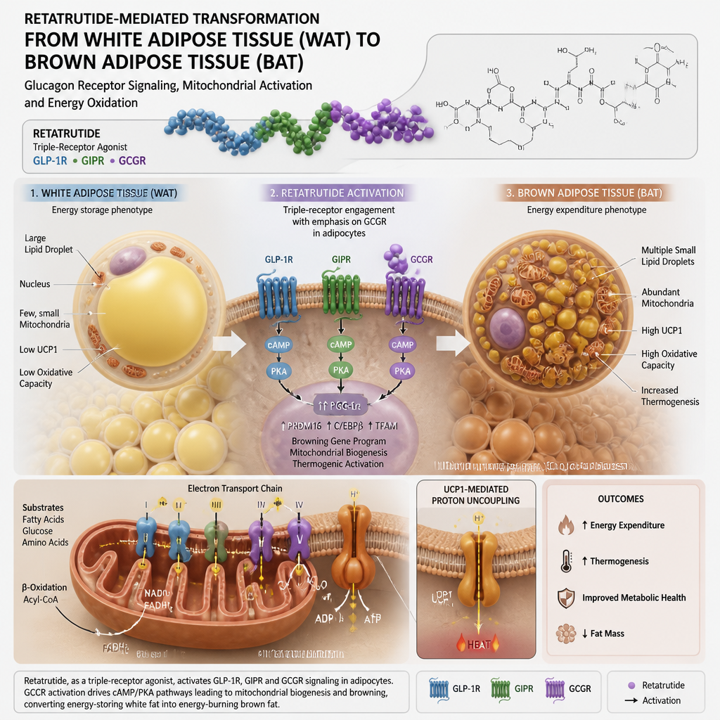
The practical implication for research laboratories is that retatrutide enables WAT browning experiments that test all three receptor contributions in a single compound series, rather than requiring parallel experiments with separate mono-agonist tools. This is especially useful when studying the interaction between lipid oxidation and thermogenesis, since the glucagon component drives fat oxidation while the GLP-1R component orchestrates the central thermogenic relay.
Pro tip: When using retatrutide in WAT browning experiments, include a glucagon receptor-selective antagonist arm in your design. This lets you isolate the fat oxidation and thermogenic contribution of the GCGR component from the GLP-1R/GIPR browning signal, producing mechanistic data that is far more publication-ready than a simple agonist-versus-vehicle comparison.
Designing In Vitro WAT-to-BAT Conversion Assays
The quality of a WAT browning assay is determined by three decisions made before any compound touches a cell: the cell model chosen, the browning readouts measured, and the compound preparation quality used. All three can independently invalidate an otherwise rigorous experimental design.
Cell Model Selection for Browning Studies
Primary stromal-vascular fraction (SVF) cells isolated from murine inguinal WAT remain the gold standard for adipose browning assays, because they retain the full complement of beige-competent precursor cells and endogenous receptor expression. 3T3-L1 cells are widely used but require validation of thermogenic gene inducibility before use in browning protocols; a significant fraction of 3T3-L1 sublines have lost efficient UCP1 induction capacity and will not show browning responses regardless of compound treatment. Human primary adipose-derived stem cells (ADSCs) are increasingly used for translational relevance but require longer differentiation timelines and higher biological replicate numbers.
Readout Selection: What Actually Confirms Browning
A common mistake in WAT browning assays is relying on UCP1 protein or mRNA alone as the sole browning confirmation. In practice, compounds that induce partial or artifactual UCP1 expression without a corresponding increase in mitochondrial biogenesis, oxygen consumption rate (OCR), and thermogenic marker co-expression (PGC-1alpha, PRDM16, CIDEA) produce irreproducible and misleading results. The minimum credible readout set for a browning assay is: UCP1 expression, OCR under uncoupled conditions via Seahorse or equivalent, and mitochondrial DNA copy number.
Dose-Response Protocol Considerations
Adipocyte browning assays with peptide compounds must account for receptor desensitization kinetics. Continuous high-dose exposure of GLP-1R in particular leads to rapid receptor internalization, which compresses the apparent potency window and produces flat or bell-shaped dose-response curves that are easy to misread. Pulsatile treatment protocols, where compound is refreshed at defined intervals rather than added once at the start of differentiation, more faithfully replicate physiological receptor engagement and produce cleaner, wider dose-response windows.
Comparison of Peptide Compound Strategies
Not all peptide browning strategies produce equivalent mechanistic data. The choice between single, dual, and triple receptor agonist tools shapes what you can and cannot conclude from any given experiment. The table below summarizes the three primary compound strategy categories used in current WAT BAT conversion peptide research.
| Compound Strategy | Receptor Targets | Key Research Advantage | Primary Limitation in Browning Studies |
|---|---|---|---|
| Single-axis GLP-1R agonist (e.g., exendin-4) | GLP-1R only | Well-characterized receptor pharmacology; decades of published reference data; useful for isolating GLP-1R-specific thermogenic signaling | Misses glucagon receptor fat oxidation contribution; central browning relay requires intact hypothalamic signaling absent in most in vitro systems |
| Dual GLP-1R/GIPR agonist | GLP-1R + GIPR | Synergistic cAMP elevation in adipocytes; captures the interaction between glucose-dependent insulinotropic and incretin axes; relevant for type 2 diabetes metabolic phenotyping | Excludes direct glucagon receptor-mediated lipid oxidation and thermogenesis; incomplete model of triple-agonist metabolic effects |
| Triple-receptor agonist (Retatrutide) | GLP-1R + GIPR + GCGR | Simultaneous activation of all three axes enabling fully integrated thermogenic reprogramming studies; most complete adipose multi-omic remodeling profile in published preclinical literature | Receptor contribution deconvolution requires selective antagonist co-treatments; compound preparation quality is critical since impurities at any of three receptor sites confound interpretation |
Why Compound Quality Determines Research Validity
In adipose browning research, compound purity is not a procurement preference, it is a methodological variable. A peptide compound below 99% purity introduces truncated sequences, oxidized residues, and related impurities that can bind target receptors with partial agonism, act as competitive antagonists, or engage off-target receptors entirely. In a browning assay measuring UCP1 induction across a six-point dose-response curve, even a small contaminating fragment with partial GLP-1R binding can produce a shifted EC50 that appears to be a genuine compound potency result but is actually an artifact of preparation quality.
For metabolic research compound procurement, the documentation trail matters as much as the purity number. Batch traceability, independent third-party laboratory verification, and chain-of-custody records are what allow researchers to attribute experimental results to the compound itself rather than to lot-to-lot variability. This is especially true for multi-receptor agonists like retatrutide, where the receptor engagement profile depends on the structural integrity of a molecule binding at three distinct receptor sites.
Pepura Labs supplies research-grade retatrutide as a lyophilized compound with purity verified at 99% or above by independent Canadian laboratories, with full batch traceability and chain-of-custody documentation. For adipose browning research teams running assays where receptor-specific mechanistic conclusions are required, this level of verification is what makes cross-batch and cross-study comparisons statistically defensible. Rapid Xpresspost delivery across Canada also means research timelines are not disrupted by multi-week international shipping delays common with overseas suppliers.
In practice, the difference between a research supplier that provides a certificate of analysis generated in-house and one that provides third-party verified documentation with full chain of custody is the difference between data you can publish and data you have to re-run. The cost difference between research-grade and sub-grade compounds is never recovered once an assay has to be repeated from scratch.
Frequently Asked Questions
What is the difference between WAT browning and beige adipocyte induction in research contexts?
WAT browning is the broader phenotypic description of white adipose tissue acquiring thermogenic characteristics, including UCP1 expression, multilocular lipid droplet morphology, and increased mitochondrial density. Beige adipocyte induction is the cellular mechanism that produces this phenotype: a WAT-resident precursor cell population differentiates or transdifferentiates toward a thermogenic cell identity under specific stimuli such as beta-adrenergic activation, cold exposure, or receptor agonist treatment. In experimental design, the terms are often used interchangeably, but mechanistic studies should specify whether they are measuring the induction of new beige cells from precursors or the activation of thermogenic function in existing beige cells, because the two processes involve different gene programs and respond differently to compound treatment timelines.
Why is the glucagon receptor component important in triple-agonist adipose browning studies?
The glucagon receptor drives fatty acid oxidation and thermogenesis through mechanisms distinct from GLP-1R and GIPR signaling. In studies using dual GLP-1R/GIPR agonists without glucagon receptor engagement, the full multi-omic adipose reprogramming profile seen with triple agonists is not reproduced. Specifically, the direct lipid oxidation component and the clearance of lipotoxic intermediates depend on glucagon receptor activation. For researchers studying the metabolic mechanisms underlying the weight-reduction efficacy of next-generation compounds, excluding the glucagon receptor from the experimental model produces an incomplete picture that can lead to incorrect mechanistic conclusions about which receptor axis is responsible for which effect.
What readouts should be included in a WAT-to-BAT conversion assay to produce publication-quality data?
The minimum credible set includes UCP1 expression at both the mRNA and protein level, oxygen consumption rate (OCR) under uncoupled conditions, and mitochondrial DNA copy number. Robust studies also add PGC-1alpha and PRDM16 expression as co-markers of the thermogenic program, cellular lipid droplet morphology via Oil Red O or equivalent, and where feasible, metabolomic profiling to capture lipid species changes associated with activated fat oxidation. Single-readout browning claims based on UCP1 mRNA alone are routinely questioned in peer review precisely because transient or partial UCP1 induction can occur without the full thermogenic reprogramming that characterizes true beige or brown adipocyte conversion.
How does lyophilized peptide storage affect browning assay outcomes?
Lyophilized peptides stored improperly before reconstitution degrade in ways that are not always visible from the compound's physical appearance. Moisture ingress during storage is the primary degradation pathway, converting intact peptide into truncated or oxidized sequences that bind receptors with altered affinity profiles. For adipose browning assays using sensitive thermogenic gene expression readouts, even partial compound degradation can shift dose-response curves by half a log unit or more. Best practice is to aliquot reconstituted compound into single-use volumes, avoid repeated freeze-thaw cycles, and use compounds supplied with full chain-of-custody documentation so that storage conditions during transit are verifiable, not assumed.
Is inguinal or epididymal WAT the preferred depot for browning research?
Inguinal subcutaneous WAT is the preferred depot for most WAT browning and beige adipocyte induction studies. It contains a higher proportion of beige-competent precursor cells and demonstrates more robust UCP1 induction in response to cold and pharmacological stimuli than epididymal visceral WAT. Epididymal WAT is more resistant to browning stimuli and is better suited to studies of adipose inflammation, insulin resistance, and lipid storage dysregulation. Using epididymal WAT as the primary browning model and expecting the same magnitude of UCP1 induction seen in inguinal depots is a common source of negative or inconsistent results in browning studies, not a true reflection of compound activity.
What makes retatrutide specifically useful for adipose browning research compared to other peptide tools?
Retatrutide's value as a research tool comes from its simultaneous engagement of three receptor axes in a single compound. This allows researchers to study the integrated thermogenic response that arises from combined GLP-1R, GIPR, and glucagon receptor activation without running three separate compound series. Published multi-omic profiling in preclinical models has documented a level of adipose tissue reprogramming with retatrutide that includes not only thermogenic gene induction but also metabolic restoration, anti-inflammatory pathway activation, and lipotoxic intermediate clearance. This comprehensive tissue remodeling signature makes it one of the most information-rich tools available for researchers studying the full scope of peptide-induced WAT browning.
If your laboratory is currently designing WAT-to-BAT conversion protocols or refining your compound sourcing strategy for adipose browning studies, share what assay parameters have proven most reliable in your hands. The experimental design decisions made at the protocol stage are what ultimately determine whether browning data holds up across repetitions and institutions.
References
- Multi-omic profiling of retatrutide effects on white adipose tissue reprogramming in obese mouse models
- GLP-1 receptor agonists and adipose tissue: mechanisms of thermogenesis and WAT-BAT phenotypic transitions
- Central GLP-1 receptor activation, BAT thermogenesis, and lipid clearance in lean and diet-induced obese mice
- Brown adipose tissue thermogenesis, UCP1 biology, and targeted strategies for WAT browning
- Novel regulatory mechanisms behind thermogenesis of brown and beige adipocytes: current research frontiers