
If your lab is characterizing dual or triple incretin receptor agonists, the cAMP assay for GLP-1 receptor activity is not optional background work. It is the primary functional readout that tells you whether your compound is actually engaging Gαs-coupled signalling at GLP-1R and GIPR, and at what potency. Designing that assay badly, through mismatched cell lines, inadequate phosphodiesterase inhibition, or poor normalization controls, produces EC50 values that drift between runs and obscure the signal bias that now defines the most interesting research on incretin pharmacology. This guide is written for research scientists who are moving past conceptual understanding and need specific decisions answered before their next experiment.
Table of Contents
- Quick Takeaways
- Why cAMP Is the Primary Readout for GLP-1 and GIP Receptor Agonism
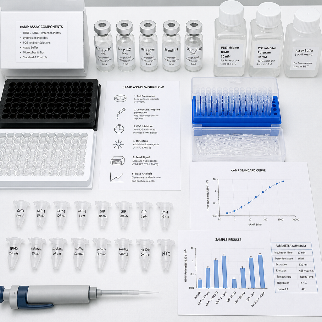
- Cell Line Selection for GLP-1R and GIPR cAMP Assays
- Detection Platform Comparison: HTRF, LANCE, EFC, and ELISA
- Assay Buffer Conditions and Phosphodiesterase Inhibition
- Signal Bias: Why cAMP Alone Does Not Tell the Full Story
- Retatrutide-Specific Assay Considerations for Triple Agonist Research
- Data Normalization and EC50 Calculation
- Frequently Asked Questions
- References
Quick Takeaways
| Key Insight | Explanation |
|---|---|
| Gαs coupling is the primary signalling axis for both GLP-1R and GIPR | Both receptors drive intracellular cAMP accumulation via Gαs; this is the mechanism underlying insulin secretion and the intended pharmacological readout for agonist characterization. |
| IBMX at 250 µM is the standard PDE inhibitor for these assays | Phosphodiesterase inhibition is mandatory to prevent cAMP degradation during incubation. Under-dosing IBMX is one of the most common causes of compressed dynamic range in GLP-1R cAMP assays. |
| HEK293 and CHO-K1 stably expressing the target receptor are both validated platforms | Each has distinct advantages. HEK293 cells support simultaneous multi-receptor characterization (GLP-1R, GIPR, GcgR); CHO-K1 offers lower endogenous GPCR background for cleaner pharmacology. |
| HTRF (CisBio cAMP Dynamic 2) is the most widely documented homogeneous format for these targets | It requires no wash steps, tolerates low cell numbers per well, and is compatible with 384-well high-throughput formats essential for peptide dose-response screens. |
| Signal bias between Gαs and β-arrestin recruitment is pharmacologically significant | A compound can show full agonism in cAMP without recruiting β-arrestin. Running cAMP assays without a parallel arrestin assay produces an incomplete and potentially misleading potency profile. |
| Receptor expression level directly affects EC50 measurement | High receptor density lowers apparent EC50 through signal amplification. For meaningful rank-ordering of peptide potency, receptor expression must be standardized across batches. |
| Normalization to the endogenous ligand is essential for cross-study comparisons | Expressing compound activity as percent of maximal native ligand response (GLP-1 7-36 amide for GLP-1R; GIP 1-42 for GIPR) is the only way to compare EC50 and Emax values across laboratories and assay formats. |
Why cAMP Is the Primary Readout for GLP-1 and GIP Receptor Agonism
Both GLP-1R and GIPR are class B G protein-coupled receptors that signal predominantly through Gαs upon agonist binding. Activation of Gαs stimulates adenylyl cyclase, which converts ATP to cyclic AMP. The resulting rise in intracellular cAMP is the immediate downstream signal that drives PKA activation, insulin secretion from pancreatic β-cells, and the broader metabolic effects that make these receptors therapeutically relevant targets. Measuring cAMP accumulation is therefore not a surrogate readout. It is the direct functional correlate of receptor activation along the primary coupling pathway.
In practice, this means that a well-designed cAMP assay for GLP-1 receptor characterization gives you two critical parameters: EC50 (potency) and Emax (intrinsic efficacy relative to the native ligand). Both are necessary. A partial agonist at GIPR with high potency may behave very differently in a physiological context than a full agonist with modest affinity, and the only way to see this distinction is through a correctly normalized cAMP concentration-response curve.
A common mistake in early assay design is treating cAMP as a binary signal rather than a graded response. Using a single compound concentration to assess "activity" produces data that cannot be translated into meaningful pharmacology. Always run a full 10-point or 12-point dilution series, starting at least two log units above your expected EC50, to capture the full sigmoidal curve.

Cell Line Selection for GLP-1R and GIPR cAMP Assays
The choice of cell line is probably the most consequential decision in assay design, and it is frequently underestimated. Two expression systems dominate the published literature on GLP-1R and GIPR cAMP pharmacology.
HEK293 Stably Expressing Target Receptors
HEK293 cells stably expressing human GLP-1R, GIPR, and GcgR are documented across multiple multi-receptor functional studies. Their advantage is that a single clonal line can be engineered to express one or more receptors at defined levels, allowing pharmacological characterization of receptor selectivity and dual or triple agonist profiling within the same experimental system. The cells transfect efficiently, grow predictably, and produce consistent cAMP responses when receptor expression is maintained under antibiotic selection pressure.
The limitation with HEK293 is endogenous GPCR expression. These cells carry a measurable panel of endogenous receptors that can contribute background cAMP signal under some stimulation conditions. For highly potent compounds, this is generally not an issue. For partial agonists or low-efficacy scaffolds, endogenous receptor crosstalk can complicate interpretation.
CHO-K1 Stably Expressing GLP-1R
CHO-K1 cells are preferred when pharmacological cleanliness is the priority. Their endogenous GPCR complement is lower than HEK293, making them the cell line of choice for compounds where signal-to-noise separation is critical. Stable CHO-K1 lines expressing human GLP-1R under an EF1A promoter with puromycin selection have been described in the literature as producing robust, reproducible cAMP responses suitable for head-to-head potency comparisons of peptide series.
A practical consideration: receptor surface expression level must be verified by flow cytometry or a binding assay before functional work begins. High and low expressing pools behave very differently in a cAMP assay, and conflating them introduces systematic error into EC50 measurements that is almost impossible to diagnose after the fact.
Pro tip: Freeze multiple early-passage vials of your verified stable line immediately after expression confirmation. Receptor expression drift under prolonged culture is real and will silently corrupt your potency data if you work from a single continuously passaged stock for more than a few weeks.
Detection Platform Comparison: HTRF, LANCE, EFC, and ELISA
Four detection formats are used in GLP-1R and GIPR cAMP assays across the research literature. Each has genuine trade-offs that depend on throughput, equipment access, and the specific questions being asked.
| Platform | Format and Principle | Best Fit for GLP-1R / GIPR Research |
|---|---|---|
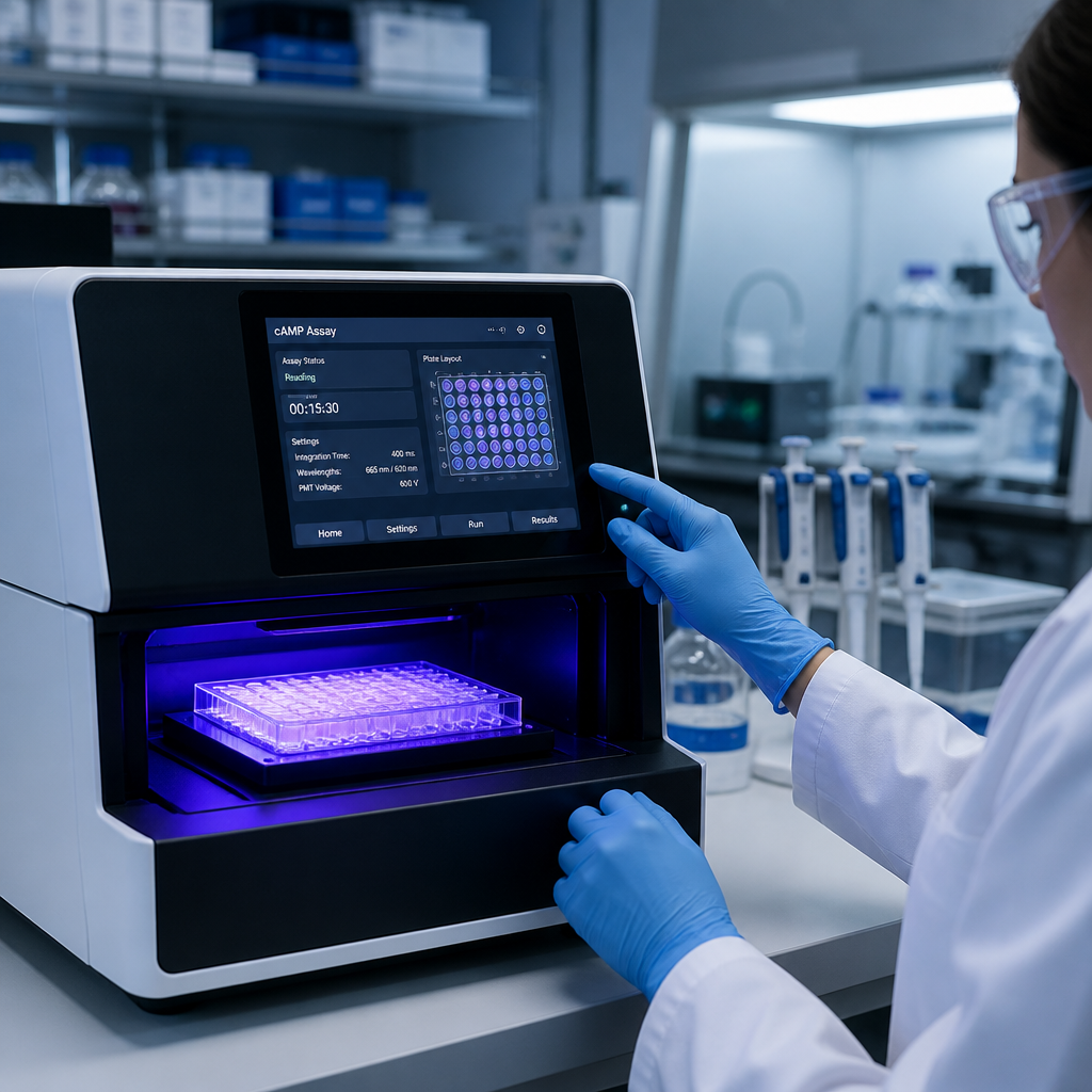
| HTRF (CisBio cAMP Dynamic 2) | Homogeneous time-resolved FRET. cAMP-d2 conjugate competes with endogenous cAMP for binding to an anti-cAMP Eu3+-Cryptate antibody. No wash steps required. Compatible with 384-well plates. | Highest documentation density for GLP-1R, GIPR, and GcgR. Preferred for multi-receptor selectivity panels and high-throughput peptide screening. Requires a TR-FRET plate reader. |
| LANCE Ultra cAMP (PerkinElmer) | Similar competitive immunoassay principle using Eu-chelate donor and ULight acceptor. Homogeneous, no wash steps. | Strong alternative when CisBio reagents are unavailable. Performance in GLP-1R assays is comparable; cross-validation against HTRF is recommended when switching between platforms mid-study. |
| EFC (Enzyme Fragment Complementation) | cAMP competes with ED-tagged cAMP for anti-cAMP antibody binding. Free ED-cAMP recombines with EA fragment to produce active β-galactosidase, detected by chemiluminescent substrate. Read on a luminescence-capable plate reader. | Well-documented for GLP-1R assays in CHO-K1 using DiscoverX cell lines. Accessible format for labs without TR-FRET readers. Longer incubation steps (up to 4 hours total) vs. HTRF. |
| cAMP ELISA (e.g., R&D Systems Parameter Kit) | Colorimetric competitive immunoassay. Cells are lysed, cAMP is measured in the lysate. Wash steps required. | Appropriate for lower-throughput mechanistic studies, including receptor characterization in PSC-derived cell lines. Lower throughput than homogeneous formats. Has been used in validated GLP-1R assay systems in academic settings. |
The practical recommendation is to use HTRF or EFC as your primary platform for dose-response screening, and reserve ELISA for endpoint confirmatory experiments or for cell types that do not plate well at the densities required for homogeneous formats.
In GLP-1R and GIPR cAMP pharmacology, the detection platform you choose determines not just assay sensitivity but the ceiling of data credibility. A result generated on a platform without cross-validation against an orthogonal method is provisional, not definitive.
Assay Buffer Conditions and Phosphodiesterase Inhibition
Phosphodiesterase (PDE) inhibition is not optional. cAMP is actively degraded by intracellular phosphodiesterases on timescales that compete directly with your assay incubation window. Without PDE inhibition, you will measure the net steady-state cAMP level rather than the peak accumulation that represents true receptor activation, and your EC50 values will be right-shifted and your Emax values compressed relative to the true pharmacology.
The standard approach in GLP-1R and GIPR cAMP assays is IBMX (3-isobutyl-1-methylxanthine) at 250 µM, prepared fresh in DMSO and diluted into assay buffer immediately before use. IBMX is a non-selective PDE inhibitor that is consistently used across published multi-receptor cAMP assay systems for incretin targets at this concentration. The assay buffer typically consists of Hanks Balanced Salt Solution with 5-20 mM HEPES at pH 7.4, supplemented with 0.1% BSA (fraction V) to reduce peptide adsorption to assay vessel surfaces. For serum-containing media formulations, 0.1% casein has also been used to prevent peptide degradation without interfering with receptor binding.
Incubation time at room temperature is typically 60 minutes for established GLP-1R and GIPR cAMP assays. Extending this to 90 minutes does not meaningfully increase signal in most formats and increases the risk of cAMP degradation even in the presence of IBMX if PDE expression is high in your chosen cell line.
Pro tip: Prepare your IBMX stock at 100 mM in DMSO and store at minus 20°C in single-use aliquots. Freeze-thaw cycling degrades IBMX activity gradually, and the effect shows up as assay-to-assay variability that is extremely difficult to trace back to the root cause without detailed reagent records.
Signal Bias: Why cAMP Alone Does Not Tell the Full Story
GLP-1R and GIPR are GPCRs, which means they can couple to multiple intracellular effectors. Gαs-driven cAMP accumulation is the primary signalling axis, but activated receptors also recruit β-arrestin 1 and β-arrestin 2, which mediate receptor internalization and initiate G protein-independent signalling. A compound that is a full agonist for cAMP can be a partial or even null agonist for β-arrestin recruitment. This is called signal bias (or biased agonism), and it is pharmacologically significant for GLP-1R research because G protein and β-arrestin pathways appear to contribute differently to downstream physiological outcomes.
Published data on GLP-1R cAMP agonism illustrates this point sharply. Native GLP-1 (7-36) and therapeutic analogues such as liraglutide produce robust β-arrestin 1 and 2 recruitment alongside cAMP accumulation. Some research compounds demonstrate full Gαs agonism with near-complete absence of β-arrestin recruitment, producing a strongly G protein-biased pharmacological profile that would not be visible from cAMP data alone.
The practical consequence for intracellular signalling peptide research is straightforward: your cAMP assay must be paired with a β-arrestin recruitment assay (PathHunter EFC or BRET-based formats are the most common) if you are generating a full pharmacological characterization. Running only cAMP will tell you that a compound activates the receptor. It will not tell you the quality or downstream selectivity of that activation.
Retatrutide-Specific Assay Considerations for Triple Agonist Research
Retatrutide (LY3437943) presents a specific assay design challenge because it is an agonist at GLP-1R, GIPR, and the glucagon receptor (GcgR). Each of these three receptors couples to Gαs and drives cAMP accumulation, which means that receptor selectivity profiling requires three parallel cAMP assays run under identical conditions, not sequential assays or assays run under different buffer conditions on different days.
Retatrutide's relative potency at each receptor differs markedly from its potency at the corresponding endogenous ligand. Published structural and pharmacological data indicates that the compound is substantially more potent at GIPR relative to native GIP, while its potency at GLP-1R and GcgR is lower than that of the respective native peptides. This pattern means that a cAMP assay run only in a GLP-1R cell line will systematically underestimate retatrutide's full pharmacological profile and could lead to misinterpretation of its mechanism of action.
For multi-receptor profiling using retatrutide or similar triple agonist scaffolds, the validated approach in published research uses HEK293 cell lines stably expressing each of the three human receptors separately, tested in parallel with identical peptide dilution series, using the HTRF cAMP Dynamic 2 format. This produces directly comparable EC50 and Emax values across all three receptors in a single experimental session.
Pro tip: When working with lyophilized retatrutide for in vitro cAMP studies, reconstitution quality directly affects assay reproducibility. Reconstitute in the solvent specified by your supplier's Certificate of Analysis, verify peptide concentration by UV absorbance or amino acid analysis before the first use, and prepare working dilutions fresh on the day of assay. Peptide adsorption to tube walls at sub-nanomolar concentrations is a genuine source of EC50 variability that is easy to underestimate and difficult to correct retroactively.
Data Normalization and EC50 Calculation
Raw cAMP assay data, whether expressed as luminescence counts, HTRF ratio, or absorbance, is meaningless without normalization. The standard approach is to express compound response as a percentage of the maximal response elicited by the endogenous reference ligand at a saturating concentration: GLP-1 (7-36) amide for GLP-1R assays, and GIP (1-42) for GIPR assays. This allows Emax to be expressed as percent of native ligand maximal response, immediately communicating whether your compound is a full agonist (Emax near 100%), a partial agonist (Emax significantly below 100%), or a super-agonist (Emax exceeding 100% relative to the native peptide).
For EC50 calculation, a four-parameter logistic (4PL) regression fit to the concentration-response data is the standard approach. Both top and bottom parameters should be allowed to float unless there is a strong prior reason to constrain them. Fixing the bottom to zero is acceptable when background signal is stable and well-characterized, but should be reported explicitly in methods sections.
Data normalization to percent stimulation using DMSO as the minimum control and the saturating native ligand as the maximum control is well-described for both GLP-1R and GIPR assay formats. When reporting GLP-1 GIP cAMP measurement data across multiple assay runs, always report within-run and between-run coefficient of variation values for your reference compound to allow readers to assess assay reproducibility. EC50 reproducibility within a factor of 2 to 3 between runs is considered acceptable for most GLP-1R and GIPR cAMP assay systems.
For researchers working with high-purity, batch-verified peptide materials, the analytical Chain of Custody for the compound tested is as important as the assay protocol itself. If your research paper or report cannot link each dataset to a specific compound lot with documented purity, you cannot claim that discordant results across experiments reflect pharmacological differences rather than compound quality differences.
Frequently Asked Questions
What concentration of IBMX should I use in a GLP-1R cAMP assay?
250 µM IBMX is the established concentration used across published GLP-1R and GIPR cAMP assay protocols in both HEK293 and CHO-K1 systems. This concentration provides broad-spectrum PDE inhibition without cell toxicity at typical assay incubation times of 60 minutes. Lower concentrations risk incomplete PDE inhibition and compressed dynamic range; higher concentrations offer no meaningful benefit and can introduce DMSO-related artefacts if the stock solution concentration requires higher solvent volumes in the assay buffer.
Can I use a transient transfection instead of a stable cell line for GLP-1R cAMP assays?
Transient transfection is viable for initial feasibility testing but is not suitable for systematic potency measurements. Receptor expression level varies substantially between transfections, which shifts apparent EC50 values and makes potency comparisons between experiments unreliable. For any dataset intended for publication or for informing compound development decisions, a stable, clonally selected cell line with documented receptor expression is required. The investment in generating a validated stable line pays back immediately in data reproducibility.
How do I account for receptor expression differences when comparing GLP-1R and GIPR potency in a dual-assay format?
Receptor expression must be matched as closely as practically possible between the two cell lines, or the apparent difference in EC50 between GLP-1R and GIPR will partly reflect expression-level differences rather than true potency differences. Quantify surface receptor expression by flow cytometry using a receptor-specific antibody or by a radioligand binding assay, and select cell pools or clones with comparable expression levels. If perfect matching is not achievable, report the receptor expression data alongside EC50 values so readers can interpret the pharmacological data in context.
What is the difference between Emax and intrinsic efficacy in the context of GIP receptor agonist signalling?
Emax is the operationally defined maximal response of a compound in a specific assay system, expressed relative to the maximal response of a reference agonist. Intrinsic efficacy (or τ in the operational model of agonism) is a system-independent property of the compound-receptor interaction. The two can diverge substantially when receptor expression is high, because signal amplification can produce full Emax responses from compounds with low intrinsic efficacy. To distinguish partial from full agonists, assays should be run in cell lines expressing receptor at or near physiological levels, or at a range of expression levels, and the data analyzed within an appropriate operational model.
Does peptide purity affect cAMP assay results, and by how much?
Yes, significantly. A peptide sample at 90% purity contains 10% impurities that may include truncated sequences, oxidized residues, or deamidated variants with different receptor binding profiles. In a cAMP assay designed to measure pharmacological potency, this can shift your apparent EC50 by a factor of two or more and alter your Emax estimate. For research-grade in vitro characterization, peptide purity of 99% or above, independently verified by HPLC and mass spectrometry with full batch documentation, is the baseline standard that allows your assay results to be meaningfully interpreted and compared across experimental sessions.
Can I run GLP-1R and GIPR cAMP assays in the same well using a multiplex format?
Not practically, because the two assays require separate cell lines expressing each receptor. You cannot simultaneously assess GLP-1R and GIPR cAMP responses in a single well using current cell-based formats without co-expression, which introduces receptor-receptor interaction artifacts. The standard approach is parallel single-receptor assays run on the same plate and on the same day with the same peptide dilution series, using common normalization controls. This is the only format that produces directly comparable EC50 and Emax values for a multi-receptor agonist such as retatrutide.
If you are currently designing a cAMP assay for GLP-1R, GIPR, or triple agonist research and have run into specific protocol challenges, share your experience in the comments. Practical field questions from working researchers help identify the gaps that no protocol paper ever covers.
We would love your feedback and any insights you would share with others. What perspective would you add?
References
- Structural and pharmacological basis of retatrutide triple agonism at GLP-1R, GIPR, and GcgR
- Biased GLP-1R and GIPR agonism: cAMP accumulation and beta-arrestin coupling characterization in recombinant cell lines
- GLP-1 receptor activation and cAMP measurement using commercial ELISA in PSC-derived cell systems
- GLP-1R cAMP assay protocol using enzyme fragment complementation in CHO-K1 cells: detailed methodology