
Picking the wrong assay endpoint in a triple-agonist peptide study does not just waste reagents. It can produce data that is mechanistically uninterpretable, force a full protocol redesign mid-experiment, or worse, lead to conclusions that do not survive peer review. For researchers running cell viability assay peptide research protocols with compounds like retatrutide, the decision between a viability readout and a receptor-level functional endpoint is one of the first choices you will make, and it shapes every downstream variable in your study. This framework will give you the reasoning and the decision criteria to make that call confidently the first time.
Table of Contents
- Quick Takeaways
- Why Endpoint Selection Defines Your Study
- Cell Viability Assays: What They Actually Measure
- Functional Endpoints in Triple-Agonist In Vitro Studies
- Retatrutide Assay Design: Receptor-Specific Considerations
- Endpoint Comparison Table
- Common Mistakes in GLP-1 GIP Glucagon Research Endpoints
- Frequently Asked Questions
- References
Quick Takeaways
| Key Insight | Explanation |
|---|---|
| Cell viability assays measure cellular health, not receptor activity | MTT, resazurin, and ATP-based assays report whether cells are alive and metabolically active. They cannot tell you whether GLP-1R, GIPR, or GCGR was actually engaged by your compound. |
| cAMP accumulation is the primary functional readout for triple-agonist GPCR studies | All three receptors targeted by retatrutide are Gs-coupled GPCRs. Intracellular cAMP generation is the most direct, quantifiable signal of agonist engagement across all three targets simultaneously. |
| Beta-arrestin recruitment catches signaling bias that cAMP misses | If your study goal includes assessing biased agonism or receptor internalization kinetics, a beta-arrestin complementation assay must run alongside the cAMP readout, not as a replacement. |
| Cell model selection determines which endpoint is valid | Overexpressed receptor cell lines are appropriate for potency ranking. Endogenous expression models such as differentiated adipocytes for GIPR or iPSC-derived hepatocytes for GCGR are required for physiologically relevant functional conclusions. |
| Peptide purity directly impacts functional endpoint reliability | At sub-nanomolar concentrations, impurities in a lower-purity peptide preparation can activate off-target pathways and create false positives in functional assays. Research-grade, 99%+ purity compounds reduce this interference significantly. |
| Viability assays are useful as a prerequisite, not a substitute | Run a viability screen first to confirm your cell model survives your peptide concentration range. Then build your functional assay stack on top of that confirmed window. |
| Lipid mobilization and glucose output are the gold-standard downstream functional endpoints | For retatrutide specifically, lipolysis in GIPR-expressing adipocytes and glucose output in GCGR-expressing hepatocytes are the most biologically grounded downstream endpoints, mirroring published preclinical data on this compound class. |
Before you open a protocol document, it is worth being explicit about why this choice matters more for triple-agonist studies than for single-target peptide work. A monoagonist study can get away with a narrower endpoint because you are querying one receptor, one signaling arm, and one downstream output. A triple-agonist compound like retatrutide engages GLP-1R, GIPR, and GCGR simultaneously, and each receptor has its own preferred signaling pathway, its own cell model requirements, and its own physiologically meaningful downstream read. Collapsing all of that complexity into a single viability number is not simplification. It is signal destruction.


Why Endpoint Selection Defines Your Study
Every assay endpoint is a filter. It selects certain biological events for measurement and ignores everything else. The question is whether the events you are selecting are the ones that actually answer your research question.
For most researchers entering triple-agonist work from a pharmaceutical or biochemical background, the default instinct is to run a viability check and treat a clean result as confirmation that the compound is "doing something." That instinct is understandable but wrong. A peptide can fully activate three GPCRs, trigger significant downstream signaling cascades, and alter gene expression profiles, all while leaving a viability readout completely unchanged, because the cells are still alive and metabolically functional. Receptor agonism at physiological concentrations does not kill cells. It changes what they do.
The inverse problem is equally common. Some researchers jump directly to a specialized downstream endpoint, such as lipolysis measurement or glucose output quantification, without first confirming that their cell model actually expresses the target receptor at a level sufficient for detection. If GCGR expression in your hepatocyte model is too low, you will get a flat dose-response curve that looks like compound failure when it is actually a model selection failure.
The hierarchy of questions to answer before choosing an endpoint
The first question is mechanistic: are you trying to confirm receptor engagement, characterize signaling pathway activation, or measure a physiologically relevant downstream biological effect? These are not the same question and they do not share the same answer.
The second question is about cell model compatibility. Receptor overexpression systems give you clean pharmacology but no physiological context. Endogenous expression models give you physiological relevance but demand more rigorous assay optimization and higher compound purity to see a signal above noise.
The third question is about your compound preparation. At concentrations below 10 nM, the functional endpoint data you collect is only as clean as the peptide you are using. Impurities in a low-purity preparation can introduce off-target effects that are indistinguishable from genuine receptor activity in a functional assay.
Pro tip: Map your research questions to endpoint categories before you design a single experiment. Write the sentence: "This study will demonstrate that [compound] [does X] in [cell model] as measured by [endpoint Y]." If you cannot complete that sentence without ambiguity, you are not ready to choose an assay.
Cell Viability Assays: What They Actually Measure
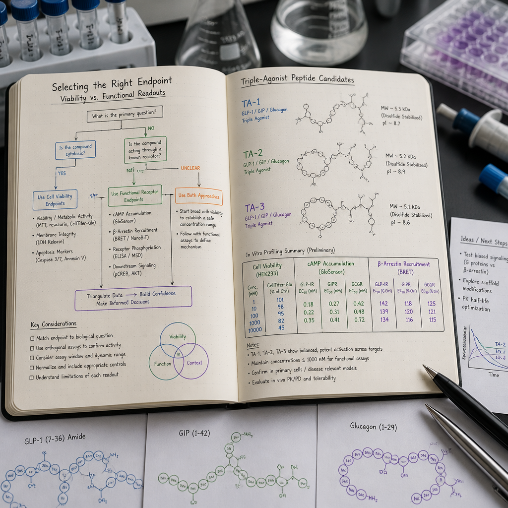
Cell viability assays measure metabolic activity as a proxy for cell survival. The three most commonly used formats, MTT/MTS tetrazolium reduction, resazurin (alamarBlue) reduction, and ATP quantification via luminescence (CellTiter-Glo), all report whether a cell is alive and mitochondrially competent. They do not report receptor occupancy, signal transduction, or downstream gene expression.
It is worth understanding the practical differences between these formats before dismissing viability assays entirely. Comparative data in the literature indicates that resazurin and ATP-based assays generally show better sensitivity than MTT-based methods, particularly at low cell numbers or when working with compounds that might interfere with the formazan reduction chemistry that MTT depends on. If you are running a viability screen to establish a safe concentration window for a peptide, the choice of viability format matters.
Where viability assays add genuine value in triple-agonist studies
Viability assays have three legitimate roles in a triple-agonist peptide study. The first is cytotoxicity screening: confirm that your compound at your intended working concentration range does not reduce cell viability below an acceptable threshold, typically 80-90% of untreated control. This step is not optional. Running a functional assay at a concentration that has already damaged your cell population will produce meaningless functional data.
The second role is quality control of your cell model. If your cells show unexpectedly low viability before compound treatment, something is wrong with your culture conditions, your passage number, or your cell density. A viability check catches those problems before you waste functional assay reagents.
The third role is in studies explicitly designed to investigate cytotoxic or cytoprotective effects. If your hypothesis is that your compound protects a cell type from a stressor, viability is the correct primary endpoint. But for mechanistic GPCR studies with retatrutide or related triple-agonist compounds, that is almost never the primary hypothesis.
Pro tip: Run your viability assay at 24 and 48 hours with your full concentration range, including concentrations above your intended working range, to build a complete safety window. Document this as a formal preliminary experiment, not a casual check, so it becomes part of your study's defensible methodology.
Functional Endpoints in Triple-Agonist In Vitro Studies
Functional endpoints for triple-agonist GPCR studies fall into three tiers, each more biologically complex and physiologically relevant than the last. Understanding which tier your study requires is the core of endpoint selection.
Tier 1: Receptor binding and cAMP accumulation
Radioreceptor binding assays confirm target engagement by measuring displacement of a labeled ligand. They give you Ki or IC50 values but no information about what happens after binding. cAMP accumulation assays are the next step: since all three receptors engaged by retatrutide, GLP-1R, GIPR, and GCGR, are Gs-coupled GPCRs, agonist binding drives adenylyl cyclase activation and intracellular cAMP production. Measuring cAMP with HTRF-based competitive immunoassays or BRET-based biosensors gives you EC50 values that are directly comparable across compounds and across receptor subtypes.
For potency ranking, early-stage characterization, and compound screening, Tier 1 is sufficient. It is fast, well-validated, and amenable to multi-well plate formats. The limitation is that it tells you nothing about signaling pathway bias or downstream cellular consequences.
Tier 2: Beta-arrestin recruitment and receptor internalization
GPCRs signal through both G proteins and beta-arrestins, and these two arms of signaling can diverge considerably depending on compound structure. For a triple-agonist compound with a complex modification profile like retatrutide, the balance between Gs-mediated cAMP signaling and beta-arrestin-mediated internalization affects receptor desensitization kinetics, duration of effect, and the cellular response to repeated dosing protocols. Beta-arrestin enzyme fragment complementation assays, such as split beta-galactosidase or split luciferase systems, provide a direct readout of this second signaling arm.
If your study is characterizing the full pharmacological profile of a triple-agonist compound or comparing signaling bias across receptor subtypes, beta-arrestin recruitment belongs in your assay panel alongside cAMP, not as an afterthought.
Tier 3: Downstream physiological endpoints
The most biologically meaningful endpoints are downstream cellular responses: lipolysis in GIPR-expressing adipocytes, glucose output in GCGR-expressing hepatocytes, and insulin secretion-related endpoints in GLP-1R-expressing pancreatic beta cell models. These require cell models with endogenous or near-endogenous receptor expression, more complex culture protocols, and longer assay timelines. But they are the endpoints that connect your in vitro data to physiological relevance.
Published characterization work on triple-agonist compounds in this class has used differentiated human adipocytes for GIPR-mediated lipolysis endpoints and iPSC-derived hepatocytes for GCGR-mediated glucose output, precisely because these models recapitulate physiological receptor expression levels and downstream signaling fidelity more accurately than simple overexpression systems.
Retatrutide Assay Design: Receptor-Specific Considerations
Retatrutide is a 39-amino-acid synthetic peptide engineered from the native GIP sequence with structural modifications that enable simultaneous activation of GIPR, GLP-1R, and GCGR. Those modifications also confer resistance to enzymatic degradation, which matters for in vitro stability but does not eliminate the need for careful handling protocols.
The key assay design challenge with retatrutide is that its three target receptors are not equally expressed across commonly used cell lines, and the relative potency at each receptor is not equal. This means that a cAMP assay run in a GLP-1R-overexpressing HEK293 cell line will give you GLP-1R activity data but will not tell you how the same compound performs at GIPR in adipocytes or GCGR in hepatocytes. A complete pharmacological characterization requires separate receptor-specific assay arms, not a single pooled readout.
Concentration range and solubility considerations
Lyophilized retatrutide should be reconstituted according to validated protocols before use. For functional cAMP assays, working concentrations typically range from picomolar to low micromolar to capture the full dose-response curve and calculate EC50 values. For viability assays run as preliminary cytotoxicity screens, testing up to a 10-fold or higher multiple of your intended functional assay concentration is standard practice.
Peptide aggregation at high concentrations can distort dose-response curves and create apparent potency losses that are actually solubility artifacts, not receptor pharmacology. Buffer composition and vehicle controls are non-negotiable parts of the protocol.
The purity requirement is not a marketing claim
At functional assay concentrations in the picomolar to nanomolar range, a 1% impurity in a peptide preparation is not negligible. That impurity, which could be a truncated sequence, a deamidated variant, or a synthesis byproduct, is present at a concentration that can independently activate receptors or interfere with detection chemistry. This is why research-grade peptide compounds for functional endpoint studies require independently verified purity documentation, not just a manufacturer's specification sheet. Batch-level certificate of analysis data with third-party verification is the minimum standard for data that will be submitted for publication or regulatory review.
In functional GPCR assays at sub-nanomolar compound concentrations, the quality of your peptide preparation is not separable from the quality of your data. They are the same thing.
Endpoint Comparison Table
The table below compares three primary endpoint approaches for triple-agonist in vitro studies across the dimensions that matter most for experimental design decisions.
| Endpoint Approach | What It Measures | Best Fit for Triple-Agonist Studies |
|---|---|---|
| Cell Viability (MTT, resazurin, CellTiter-Glo) | Metabolic activity and cell survival; reports cytotoxicity or cytoprotection | Cytotoxicity screening and concentration window validation only. Cannot confirm receptor engagement, potency, or pathway activation. Use as a prerequisite step, never as a primary mechanistic endpoint. |
| cAMP Accumulation (HTRF, BRET biosensor) | Gs-coupled receptor activation; quantifies intracellular cAMP production per receptor | Primary pharmacological endpoint for GLP-1R, GIPR, and GCGR agonism. Enables EC50 determination and cross-receptor potency comparison. Requires receptor-appropriate cell models for each target arm. High throughput compatible. |
| Downstream Physiological Endpoint (lipolysis, glucose output, insulin secretion) | Cell-type-specific downstream responses driven by receptor activation | Highest biological relevance for GIPR (lipolysis in adipocytes) and GCGR (glucose output in hepatocytes). Requires endogenous expression models, longer protocols, and higher compound purity standards. Essential for studies making physiologically grounded mechanistic claims. |
Common Mistakes in GLP-1 GIP Glucagon Research Endpoints
The most common error in this space is using a receptor overexpression system to generate potency data and then making direct physiological claims based on those numbers. Overexpression systems amplify the cAMP signal, which means your EC50 values will be lower than what you would see in a physiological expression context. This is not a problem if you are comparing compounds relative to each other in the same system. It becomes a problem if you present overexpression-derived EC50 values as predictive of in vivo potency without qualification.
The second common mistake is running all three receptor arms in the same overexpressed cell line simultaneously. If you co-express GLP-1R, GIPR, and GCGR in one cell line and treat with retatrutide, your cAMP readout is a composite of all three receptor contributions. You cannot deconvolute individual receptor contributions from that pooled signal. Use separate cell lines for each receptor arm, or use receptor-selective antagonists to isolate each contribution.
The third mistake is failing to validate your cell model before interpreting negative results. If your dose-response curve is flat, it could mean the compound is inactive, or it could mean your cells do not express the target receptor at a detectable level, your assay window is too narrow, or your compound was degraded before it reached the receptor. A flat curve is not interpretable without receptor expression verification by western blot or qPCR, a positive control compound with known activity, and a confirmed compound stability check at your experimental conditions.
The fourth mistake is treating peptide purity as a procurement detail rather than an experimental variable. In functional endpoint studies, peptide identity and purity belong in the Methods section, and batch-specific documentation should be archived with your raw data. If you cannot trace a specific data point back to a specific batch with verified composition, your study has a reproducibility gap that reviewers will find.
Pro tip: When sourcing research-grade retatrutide for functional endpoint studies, request the batch-specific certificate of analysis and confirm that purity verification was performed by an independent laboratory, not just by the supplier's in-house analytical team. For Canadian researchers, Chain of Custody documentation from a Canadian laboratory provides an additional layer of data integrity that is increasingly expected in institutional review contexts.
Frequently Asked Questions
Can a cell viability assay substitute for a functional endpoint in a retatrutide study?
No, and this substitution is a design error rather than a simplification. Cell viability assays measure whether cells are alive and metabolically active. They do not measure receptor engagement, cAMP production, or any downstream signaling event. Retatrutide at pharmacologically relevant concentrations activates GPCRs without causing cytotoxicity, so a viability readout will appear completely unchanged even when robust receptor activation is occurring. If your research question concerns the pharmacological activity of the compound, viability is not an endpoint. It is a quality control check.
What cell models are most appropriate for a triple-agonist in vitro study targeting all three receptors?
The answer depends on your study tier. For potency characterization and EC50 determination, separate receptor-overexpressing cell lines, typically HEK293-based, for each of GLP-1R, GIPR, and GCGR are the standard choice. For physiologically relevant functional endpoints, differentiated human adipocytes with endogenous GIPR expression are appropriate for lipolysis endpoints, and iPSC-derived hepatocytes with endogenous GCGR expression are appropriate for glucose output endpoints. GLP-1R-mediated endpoints in physiological models typically require pancreatic beta cell lines or primary islet preparations. None of these models is interchangeable. Select based on which receptor arm your primary hypothesis targets.
Why does peptide purity matter specifically for functional endpoint assays?
Functional endpoint assays, particularly cAMP and beta-arrestin recruitment assays, operate at compound concentrations in the picomolar to low nanomolar range. At these concentrations, a 1% impurity in a 95% purity peptide preparation is present at a concentration that can independently activate receptors, interfere with detection antibodies, or alter cell signaling in ways that confound your primary readout. Research-grade peptides verified at 99%+ purity by independent analytical laboratory methods reduce this interference. For studies generating data intended for publication or institutional reporting, batch-specific purity documentation is a methodological requirement, not an optional convenience.
How do I know when to add a beta-arrestin recruitment assay to my study panel?
Add a beta-arrestin recruitment assay when your study hypothesis includes any of the following: characterizing signaling pathway bias between Gs and beta-arrestin arms for a test compound, comparing internalization kinetics across structural analogs of a triple-agonist compound, investigating receptor desensitization after repeated dosing, or generating a pharmacological profile intended to support structure-activity relationship analysis. If your study is purely a potency screen or a single-point confirmation of receptor engagement, a cAMP assay alone is sufficient. Beta-arrestin assays add time, cost, and analytical complexity. Deploy them when the scientific question requires them, not by default.
What positive controls should I use in a retatrutide functional endpoint study?
Use a receptor-selective positive control for each receptor arm you are assaying. For GLP-1R, exendin-4 or native GLP-1(7-36) amide at a saturating concentration is a well-validated control with published EC50 values across multiple cell models. For GIPR, native GIP(1-42) serves the same function. For GCGR, native glucagon is the appropriate control. For each control, your assay should reproduce the expected maximum signal and approximate EC50 before any test compound data is considered valid. A functional assay that does not validate with a known positive control on the same plate has a protocol problem that no amount of test compound data can fix.
Is it acceptable to run all three receptor assays on the same day with the same peptide stock?
Yes, using the same reconstituted peptide stock across receptor arms on the same day is standard practice and preferable to preparing separate stocks, because it eliminates inter-stock variability as a confounding factor. Prepare your reconstituted stock, confirm concentration by absorbance or established quantification method, aliquot into single-use volumes to avoid freeze-thaw degradation, and document the preparation date and batch number. If you are running assays across multiple days, use fresh aliquots each day rather than refreezing and rethawing the same tube. Peptide stability under your specific storage and handling conditions should be confirmed as part of your preliminary assay development.
If you are currently designing a triple-agonist in vitro study and have run into endpoint selection challenges or peptide sourcing issues specific to your cell model, share your protocol context in the comments. The more specific the question, the more useful the discussion becomes for the broader research community.
References
- Published characterization of a triple GLP-1/GIP/glucagon receptor agonist with functional endpoint data in adipocytes and hepatocytes (Cell Metabolism)
- Structural basis of simultaneous GLP-1R, GIPR, and GCGR activation by retatrutide and cAMP signaling potency data (Cell Discovery, Nature Publishing Group)
- Comparative sensitivity of real-time and endpoint cell viability assay formats including resazurin, CellTiter-Glo, and MTT (ScienceDirect)
- Beta-arrestin recruitment assay methodology for GPCR ligand characterization and detection of biased agonism (PubMed Central)