
Research
How to Reconstitute Retatrutide for In Vitro Research
September 1, 2026
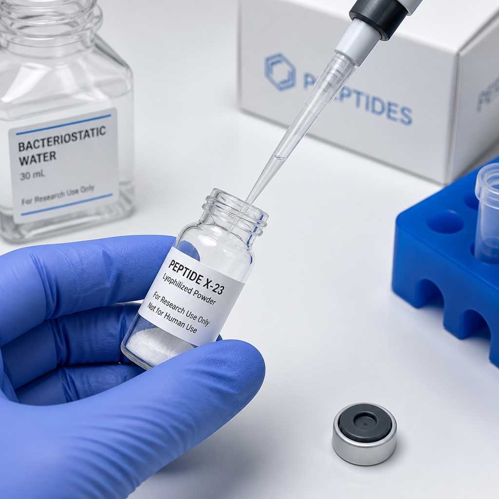
Most reconstitution errors happen in the first thirty seconds. A researcher draws the bacteriostatic water, injects it straight into the lyophilized cake with force, and inadvertently shears the peptide structure before any measurement is taken. With Retatrutide, a 39-amino-acid lipid-conjugated triple-receptor agonist, that kind of handling mistake is not recoverable. This step-by-step protocol covers how to reconstitute Retatrutide correctly for in vitro research, from equipment selection through post-reconstitution storage, so every microgram of your research-grade compound reaches the assay intact.
Table of Contents
- Quick Takeaways
- What Is Retatrutide and Why Reconstitution Matters
- Equipment and Materials Checklist
- Step-by-Step Reconstitution Protocol This protocol reflects standard aseptic technique for lyophilized peptide reconstitution. Each step is non-negotiable for maintaining sterility and structural integrity of the compound. Step 1: Prepare the Work Area Wipe down the laminar flow hood surface with 70% isopropyl alcohol and allow it to dry completely. Put on sterile gloves. Do not touch the interior of the hood surface after cleaning. Lay out all materials within arm's reach so no reaching across the field is necessary during reconstitution. Step 2: Swab All Septums Using a fresh alcohol swab, wipe the rubber septum of both the Retatrutide vial and the bacteriostatic water vial. Allow them to air-dry for 15-20 seconds. Do not blow on them to speed drying. The alcohol film must evaporate fully before needle penetration to avoid introducing alcohol into the vial. Step 3: Draw the Bacteriostatic Water Insert the syringe needle through the septum of the bacteriostatic water vial and draw the calculated volume of diluent. Work at a slight angle to minimize coring of the rubber septum. Keep the needle tip submerged in the liquid when drawing to avoid pulling in air bubbles. The common reconstitution volumes used in research are: 2 mL added to a 20 mg vial yields a 10 mg/mL working concentration, and 2 mL added to a 40 mg vial yields a 20 mg/mL concentration. For a lower concentration stock, 3 mL added to a 40 mg vial yields approximately 13 mg/mL. Step 4: Inject Along the Inner Vial Wall This is the most critical manual step. Insert the needle through the septum of the Retatrutide vial and angle the needle tip so it points toward the inner glass wall, not downward into the lyophilized cake. Depress the plunger slowly and steadily, letting the bacteriostatic water run down the inside wall of the vial and pool at the base rather than hitting the powder directly. A slow, controlled injection over 10-15 seconds is the target pace. Step 5: Swirl Gently Until Dissolved Once all the diluent is added, remove the needle and cap the syringe safely. Hold the vial between your fingers and rotate it in slow circles. Do not invert rapidly or shake. The lyophilized plug should dissolve within 30-90 seconds in most cases. If any visible particles remain after 2 minutes, continue gentle swirling. Do not force dissolution by shaking. Step 6: Inspect the Solution Hold the vial up to a light source and examine the liquid. A properly reconstituted Retatrutide solution should appear clear and colorless, free of visible particulate matter or cloudiness. Slight foaming at the surface immediately after mixing is normal and will dissipate. If the solution is cloudy, has floating particulates, or shows any color other than clear after the foam dissipates, do not use it. Step 7: Label and Store Immediately label the vial with the compound name, calculated concentration, date of reconstitution, and the initials of the preparer. Store the reconstituted vial at 2-8°C in a refrigerator. Keep it protected from direct light. Do not freeze the reconstituted liquid. Pro tip: If your research protocol requires multiple aliquots over several weeks, consider preparing small-volume single-use aliquots at reconstitution time and freezing them at -20°C before any have been punctured. Each frozen aliquot is then thawed once and used, eliminating repeat freeze-thaw cycles on the main stock. Once an aliquot is thawed, it is refrigerated and used within the same window as standard reconstituted stock. Calculating Your Working Concentration
- Solvent Selection: Bacteriostatic Water vs. Alternatives
- Storage and Stability After Reconstitution
- Common Mistakes and How to Avoid Them The most frequent errors in peptide reconstitution protocols cluster around three areas: solvent injection technique, concentration calculation, and post-reconstitution storage. Understanding why each error matters helps build habits that prevent it. Injecting Directly Into the Lyophilized Cake Directing the stream of bacteriostatic water straight down onto the powder plug causes immediate foaming and mechanical disruption of the peptide structure. The solution may appear to dissolve, but the shear forces from direct injection can cause aggregation or partial denaturation that is not visible to the naked eye. Always angle the needle toward the inner glass wall and let gravity and capillary action do the mixing work. Shaking Instead of Swirling Shaking introduces air bubbles and applies repeated mechanical force to the peptide chains. Swirling, by contrast, relies on gentle fluid movement. This matters specifically for a lipid-conjugated peptide like Retatrutide because the fatty acid moiety can form micelles or aggregates when air is incorporated under mechanical stress. Skipping the Visual Inspection Step Researchers under time pressure frequently skip the post-reconstitution clarity check. This is a mistake. A contaminated or degraded solution that passes into an assay generates confounding data, and the contamination event may not be traceable after the fact. Visual inspection takes under 30 seconds and is the last line of defense before the compound enters experimental conditions. Using Non-Verified Reagent-Grade Material In vitro research depends on knowing exactly what is in the vial. Retatrutide sourced without independent third-party verification or without batch documentation introduces variables that cannot be controlled for in experimental design. Pepura Labs provides independently verified 99%+ purity Retatrutide with full Chain of Custody documentation precisely because undocumented material is scientifically unreliable, regardless of what the supplier claims. Reproducibility requires verified inputs. Frequently Asked Questions
- References
Quick Takeaways
| Key Insight | Explanation |
|---|---|
| Bacteriostatic water is the preferred diluent | The 0.9% benzyl alcohol preservative inhibits microbial growth and extends reconstituted peptide usability to 28-30 days at 2-8°C, versus 24-48 hours with plain sterile water. |
| Never inject directly onto the lyophilized cake | Direct high-pressure injection causes foaming and mechanical degradation. Aim the stream down the inner wall of the vial so the liquid rolls gently over the powder. |
| Swirl, never shake | Vigorous shaking introduces air bubbles and can denature the peptide. Gentle swirling until fully dissolved is the correct technique. |
| Verify solution clarity before use | A properly reconstituted Retatrutide solution should be clear and colorless. Cloudiness, particulate matter, or discoloration are signs of contamination or degradation. |
| Store lyophilized powder at -20°C long-term | Sealed, desiccated lyophilized Retatrutide stored frozen at -20°C or colder is generally stable for 12 months or longer. Once reconstituted, switch to refrigerator storage only. |
| Concentration math must be done before you reconstitute | Decide your working concentration before adding diluent, not after. Adding the wrong volume of bacteriostatic water is the most common source of dosing error in in vitro assays. |
| Batch traceability matters for reproducibility | In vitro research depends on documented, verified material. Using a supplier that provides Chain of Custody documentation and independent lab verification protects the integrity of your data. |
What Is Retatrutide and Why Reconstitution Matters
Retatrutide (CAS 2381089-83-2) is a synthetic 39-amino-acid, lipid-conjugated peptide classified as a unimolecular GIP/GLP-1/glucagon triple receptor agonist. Its sequence incorporates non-canonical alpha-aminoisobutyric acid (Aib) residues and carries a lysine-conjugated fatty-diacid moiety, which is precisely what makes it structurally sensitive during handling. Research-grade Retatrutide is supplied as a lyophilized solid at 99%+ HPLC purity, and that purity figure means nothing if the reconstitution step introduces contamination, degrades the peptide through mechanical shear, or results in an incorrectly calculated working concentration.
For in vitro applications, reconstitution quality directly affects assay reproducibility. A degraded or contaminated solution does not just waste the vial. It produces data that cannot be replicated, which is the worst possible outcome in a research setting. Every component of the protocol below exists for a specific reason, and each step connects back to protecting the structural integrity and biological activity of the compound.
Equipment and Materials Checklist
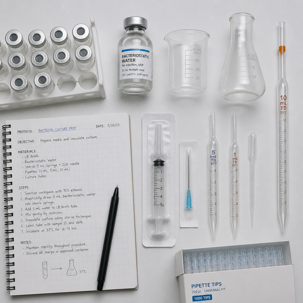
Before opening the vial, have everything ready on a clean work surface inside a laminar flow hood or equivalent sterile environment. Interrupting the protocol mid-step to locate a missing syringe is how contamination enters.
Required Supplies
- Lyophilized Retatrutide vial (research-grade, 99%+ purity, with batch documentation)
- Bacteriostatic water for injection (BAC water, 0.9% benzyl alcohol)
- Sterile insulin syringes or low-dead-volume syringes (1 mL, graduated in 0.01 mL increments)
- Alcohol swabs (70% isopropyl alcohol)
- Sterile gloves and lab coat
- Laminar flow hood or clean bench
- Refrigerator-safe amber or light-protective vial storage
- Permanent marker and label tape for concentration and date notation
Pro tip: Use a low-dead-volume insulin syringe rather than a standard 3 mL syringe when adding bacteriostatic water. Residual volume trapped in standard syringe dead space becomes significant at the small volumes used in peptide reconstitution, and it throws off your actual concentration.
What to Verify Before You Begin
Check the vial's appearance before opening. The lyophilized cake should be a white to off-white powder or plug, firmly seated in the vial. Any sign of moisture, caking that looks wet, or visible discoloration indicates that cold-chain integrity may have been compromised. For Pepura Labs vials, the accompanying Chain of Custody documentation and independent Canadian lab verification gives you a baseline reference point for what the compound should look like and what purity was confirmed at batch release. If the physical appearance does not match, do not proceed.
Also confirm that you are working with the correct vial size for your intended concentration. Common Pepura Labs research vial sizes are 10 mg and 20 mg. Know which you have before you calculate diluent volume.
Step-by-Step Reconstitution Protocol
This protocol reflects standard aseptic technique for lyophilized peptide reconstitution. Each step is non-negotiable for maintaining sterility and structural integrity of the compound.
Step 1: Prepare the Work Area
Wipe down the laminar flow hood surface with 70% isopropyl alcohol and allow it to dry completely. Put on sterile gloves. Do not touch the interior of the hood surface after cleaning. Lay out all materials within arm's reach so no reaching across the field is necessary during reconstitution.
Step 2: Swab All Septums
Using a fresh alcohol swab, wipe the rubber septum of both the Retatrutide vial and the bacteriostatic water vial. Allow them to air-dry for 15-20 seconds. Do not blow on them to speed drying. The alcohol film must evaporate fully before needle penetration to avoid introducing alcohol into the vial.
Step 3: Draw the Bacteriostatic Water
Insert the syringe needle through the septum of the bacteriostatic water vial and draw the calculated volume of diluent. Work at a slight angle to minimize coring of the rubber septum. Keep the needle tip submerged in the liquid when drawing to avoid pulling in air bubbles. The common reconstitution volumes used in research are: 2 mL added to a 20 mg vial yields a 10 mg/mL working concentration, and 2 mL added to a 40 mg vial yields a 20 mg/mL concentration. For a lower concentration stock, 3 mL added to a 40 mg vial yields approximately 13 mg/mL.
Step 4: Inject Along the Inner Vial Wall
This is the most critical manual step. Insert the needle through the septum of the Retatrutide vial and angle the needle tip so it points toward the inner glass wall, not downward into the lyophilized cake. Depress the plunger slowly and steadily, letting the bacteriostatic water run down the inside wall of the vial and pool at the base rather than hitting the powder directly. A slow, controlled injection over 10-15 seconds is the target pace.
Step 5: Swirl Gently Until Dissolved
Once all the diluent is added, remove the needle and cap the syringe safely. Hold the vial between your fingers and rotate it in slow circles. Do not invert rapidly or shake. The lyophilized plug should dissolve within 30-90 seconds in most cases. If any visible particles remain after 2 minutes, continue gentle swirling. Do not force dissolution by shaking.
Step 6: Inspect the Solution
Hold the vial up to a light source and examine the liquid. A properly reconstituted Retatrutide solution should appear clear and colorless, free of visible particulate matter or cloudiness. Slight foaming at the surface immediately after mixing is normal and will dissipate. If the solution is cloudy, has floating particulates, or shows any color other than clear after the foam dissipates, do not use it.
Step 7: Label and Store
Immediately label the vial with the compound name, calculated concentration, date of reconstitution, and the initials of the preparer. Store the reconstituted vial at 2-8°C in a refrigerator. Keep it protected from direct light. Do not freeze the reconstituted liquid.
Pro tip: If your research protocol requires multiple aliquots over several weeks, consider preparing small-volume single-use aliquots at reconstitution time and freezing them at -20°C before any have been punctured. Each frozen aliquot is then thawed once and used, eliminating repeat freeze-thaw cycles on the main stock. Once an aliquot is thawed, it is refrigerated and used within the same window as standard reconstituted stock.
Calculating Your Working Concentration
The formula is straightforward: divide the total mass of peptide in the vial by the volume of diluent added. The result is your stock concentration in the units you used for mass and volume.
For a 10 mg vial with 1 mL bacteriostatic water: 10 mg / 1 mL = 10 mg/mL (10,000 mcg/mL). For a 10 mg vial with 2 mL bacteriostatic water: 10 mg / 2 mL = 5 mg/mL (5,000 mcg/mL). To calculate volume for a specific dose from that stock, divide the target dose by the stock concentration. If your in vitro assay requires 0.1 mg per well and your stock is 5 mg/mL, you need 0.02 mL (20 microliters) per well.
A common mistake is conflating milligrams with micrograms mid-calculation. Write out units explicitly at every step. Errors at the calculation stage are invisible at the bench and only become apparent when results are inconsistent across replicates.
The concentration calculation must happen before you add diluent, not after. Once you have added the wrong volume of bacteriostatic water, your only options are to discard the vial or accept imprecise working concentrations. There is no correcting it retroactively.
Solvent Selection: Bacteriostatic Water vs. Alternatives
For research-grade Retatrutide reconstitution intended for in vitro multi-use, bacteriostatic water is the correct choice. The 0.9% benzyl alcohol content provides antimicrobial protection between uses, extending the usable window of the reconstituted solution to 28-30 days when stored properly at 2-8°C. Sterile water for injection (SWFI), which contains no preservative, must be used within 24-48 hours of reconstitution and introduces substantially higher contamination risk if the vial is re-entered multiple times.
Some researchers ask about DMSO (dimethyl sulfoxide) as a solvent. For in vitro cell-based assays, DMSO at concentrations above 0.1% can itself affect cell viability and confound results. Unless the specific assay protocol explicitly requires DMSO as a co-solvent for solubility reasons and controls are built in, bacteriostatic water remains the standard.
Comparison of Reconstitution Solvents for In Vitro Retatrutide Research
| Solvent | Usable Window After Reconstitution | Best Use Case |
|---|---|---|
| Bacteriostatic Water (0.9% benzyl alcohol) | 28-30 days at 2-8°C (multi-use vials) | Standard in vitro research protocols requiring repeated aliquoting from the same vial over several weeks |
| Sterile Water for Injection (SWFI) | 24-48 hours (single-use only) | Single-use assays where the entire vial will be used immediately; or where benzyl alcohol must be excluded from the assay design |
| Acetic Acid (0.1-1%) in Sterile Water | Variable; generally shorter shelf life, requires specific protocol validation | Some peptides with poor aqueous solubility that require a mildly acidic environment; not a default for Retatrutide and requires validation before use |
Storage and Stability After Reconstitution
Lyophilized Retatrutide in its sealed vial, stored desiccated and frozen at -20°C or below, is generally stable for 12 months or longer when protected from light and moisture. The powder form is significantly more stable than the reconstituted liquid, which is why best practice is to reconstitute only what will be used within the relevant time window rather than reconstituting an entire stock at once.
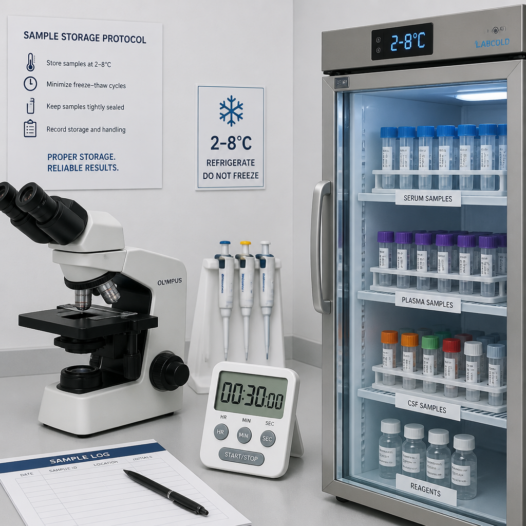
Once reconstituted with bacteriostatic water, store the vial at 2-8°C, away from direct light, and do not freeze it. Freezing reconstituted peptide can cause aggregation and concentration inconsistency when thawed. The exception is the deliberate single-use aliquot strategy described in the protocol steps above, where individual aliquots are frozen before any needle re-entry, which is a different practice from refreezing a partially used vial.
Protecting Against Repeat Freeze-Thaw Cycles
Each freeze-thaw cycle a reconstituted peptide solution goes through introduces the possibility of ice crystal formation, which can disrupt the peptide's lipid conjugate and alter its structural characteristics. For a compound like Retatrutide, where the fatty-diacid moiety on the lysine residue is central to its receptor interactions, protecting that structural feature during storage is part of maintaining assay relevance. In practice, this means that if you anticipate needing the same batch of Retatrutide across multiple experiment days, pre-aliquoting at reconstitution into volumes sized for one day's use is the most reliable approach.
Common Mistakes and How to Avoid Them
The most frequent errors in peptide reconstitution protocols cluster around three areas: solvent injection technique, concentration calculation, and post-reconstitution storage. Understanding why each error matters helps build habits that prevent it.
Injecting Directly Into the Lyophilized Cake
Directing the stream of bacteriostatic water straight down onto the powder plug causes immediate foaming and mechanical disruption of the peptide structure. The solution may appear to dissolve, but the shear forces from direct injection can cause aggregation or partial denaturation that is not visible to the naked eye. Always angle the needle toward the inner glass wall and let gravity and capillary action do the mixing work.
Shaking Instead of Swirling
Shaking introduces air bubbles and applies repeated mechanical force to the peptide chains. Swirling, by contrast, relies on gentle fluid movement. This matters specifically for a lipid-conjugated peptide like Retatrutide because the fatty acid moiety can form micelles or aggregates when air is incorporated under mechanical stress.
Skipping the Visual Inspection Step
Researchers under time pressure frequently skip the post-reconstitution clarity check. This is a mistake. A contaminated or degraded solution that passes into an assay generates confounding data, and the contamination event may not be traceable after the fact. Visual inspection takes under 30 seconds and is the last line of defense before the compound enters experimental conditions.
Using Non-Verified Reagent-Grade Material
In vitro research depends on knowing exactly what is in the vial. Retatrutide sourced without independent third-party verification or without batch documentation introduces variables that cannot be controlled for in experimental design. Pepura Labs provides independently verified 99%+ purity Retatrutide with full Chain of Custody documentation precisely because undocumented material is scientifically unreliable, regardless of what the supplier claims. Reproducibility requires verified inputs.
Frequently Asked Questions
What is the correct volume of bacteriostatic water to use for a 10 mg Retatrutide vial?
For a 10 mg vial, adding 1 mL of bacteriostatic water yields a 10 mg/mL stock concentration. Adding 2 mL yields a 5 mg/mL stock. The right choice depends entirely on your assay's required working concentration. Calculate what your protocol needs before opening the vial so you add the correct volume on the first attempt.
Can I use sterile saline instead of bacteriostatic water to reconstitute Retatrutide?
Sterile saline (0.9% NaCl) is not the preferred diluent for peptide reconstitution intended for multi-use. It contains no antimicrobial preservative, meaning bacterial growth is uninhibited after the first vial entry. If your assay requires isotonic conditions and benzyl alcohol must be excluded, use sterile water for injection instead and treat the vial as single-use, with a 24-48 hour window after reconstitution.
How long does reconstituted Retatrutide remain stable?
When reconstituted with bacteriostatic water and stored at 2-8°C in a refrigerator protected from direct light, reconstituted Retatrutide is generally stable for 28-30 days. If reconstituted with plain sterile water without a benzyl alcohol preservative, use within 24-48 hours and under strict aseptic conditions. Do not refreeze a partially used reconstituted vial.
Why does my reconstituted Retatrutide look slightly foamy right after mixing?
Slight foaming immediately after adding bacteriostatic water and swirling is a normal finding during peptide reconstitution. The foam should dissipate within a few minutes as you allow the vial to sit undisturbed. If significant foaming persists beyond 5 minutes, or if the solution remains turbid or cloudy after the foam clears, that is a sign of a problem and the solution should not be used.
Does Retatrutide need to be reconstituted differently for in vitro use compared to other research protocols?
The core reconstitution technique is consistent: bacteriostatic water, wall-injection method, gentle swirling, clarity check. However, for in vitro cell-based assays, pay close attention to the final benzyl alcohol concentration your cells will be exposed to at working dilution. At high working concentrations with minimal dilution into culture media, the benzyl alcohol from the bacteriostatic water can affect cell viability. Always dilute your stock sufficiently in the assay buffer or media to reduce benzyl alcohol to a non-cytotoxic level, and include a vehicle control in your experimental design.
What should I do if the lyophilized Retatrutide powder looks discolored or clumped before reconstitution?
Do not proceed with reconstitution. Discoloration, wet-looking caking, or an unusual odor from a lyophilized peptide vial suggests that moisture infiltration or temperature excursion has occurred during storage or shipping. Contact your supplier and reference the batch documentation and Chain of Custody records to determine whether the product was handled within specification. A reputable research supplier will have documentation to support or refute an integrity claim at the batch level.
If you are using Retatrutide from Pepura Labs in your research, we would like to hear how your reconstitution protocol is working and whether there are specific in vitro applications you would like us to address in future technical guides.
We would love your feedback and any insights you would share with others. What perspective would you add?
References
- Physician-authored guide covering safe Retatrutide mixing process and post-reconstitution storage principles
- Retatrutide researcher dosing guide covering reconstitution volumes, step-by-step protocol, and storage guidelines
- Technical reference on Retatrutide peptide structure, CAS number, purity classification, and lyophilized storage conditions
- Complete guide to bacteriostatic water use in lyophilized peptide reconstitution, sterility, and best practices
- Retatrutide protocol reference covering reconstitution math, storage matrix, and handling considerations for researchers