
Research
GLP-1 Receptor Agonist Mechanisms in Retatrutide
August 29, 2026
Most researchers studying incretin biology enter retatrutide through the lens of weight loss pharmacology, which is understandable but limiting. What makes retatrutide scientifically distinctive is not simply that it activates three receptors, but how it activates them, and specifically, what the GLP-1 receptor agonist component contributes at the molecular level in ways that differ from a classical GLP-1 monotherapy. For in vitro researchers, understanding this distinction is not academic. It shapes every experimental design decision, from cell line selection to assay endpoint choice to compound handling. This article breaks down the GLP-1 receptor mechanism in retatrutide, examines what the structural biology tells us about receptor engagement, and addresses what that means practically for bench-level research work with this compound.
Table of Contents
- Quick Takeaways
- Retatrutide as a Triple Receptor Agonist: Where GLP-1R Fits
- The GLP-1R Signaling Pathway: cAMP, PKA, and Downstream Cascades
- Structural Basis of GLP-1R Engagement in Retatrutide
- In Vitro Research Implications: Cell Models, Assays, and Endpoints
- Comparing GLP-1R Engagement Across Single, Dual, and Triple Agonists
- Compound Handling and Purity Considerations for GLP-1R Research
- Frequently Asked Questions
- References
Quick Takeaways
| Key Insight | Explanation |
|---|---|
| Retatrutide is a GLP-1/GIP/glucagon triple agonist | It simultaneously activates GLP-1R, GIPR, and GCGR, meaning in vitro results cannot be attributed to GLP-1R alone without receptor-specific controls. |
| GLP-1R agonism drives appetite and glycemic signaling | The GLP-1R component contributes hypothalamic appetite suppression, slowed gastric emptying effects, and glucose-stimulated insulin potentiation. |
| Retatrutide has approximately 2.5x lower GLP-1R potency than native GLP-1 | This matters for EC50-based in vitro assay design. Dose-response curves will shift relative to GLP-1 or semaglutide reference standards. |
| cAMP production is the primary in vitro readout for GLP-1R activation | GLP-1R couples to Gs proteins, activating adenylate cyclase and generating cAMP, which is quantifiable via HTRF, LANCE, or HitHunter assay formats. |
| ECL1 conformation differences distinguish GLP-1R from GIPR and GCGR engagement | Cryo-EM structural data shows receptor-specific conformations, particularly in extracellular loop 1, that enable retatrutide's multi-receptor activity. |
| C20 fatty diacid moiety extends half-life via albumin binding | This structural feature, attached at lysine-20, gives retatrutide a half-life of approximately six days, which directly affects in vitro washout and repeat-dosing experiment design. |
| Purity grade determines signal reliability in GLP-1R assays | Sub-99% purity lots introduce receptor-binding impurities that confound cAMP readouts. Research-grade verification is not optional for mechanistic work. |
Retatrutide as a Triple Receptor Agonist: Where GLP-1R Fits
Retatrutide (LY3437943) is a 39 amino-acid synthetic peptide engineered from a GIP peptide backbone. It is conjugated to a C20 fatty diacid moiety via a gamma-glutamic acid spacer at lysine-20, which mediates albumin binding and extends its pharmacokinetic half-life to approximately six days in preclinical research models. This structural design places it in a different class than earlier GLP-1 monotherapies.
The compound acts as an agonist at three distinct receptors: the glucagon-like peptide-1 receptor (GLP-1R), the glucose-dependent insulinotropic polypeptide receptor (GIPR), and the glucagon receptor (GCGR). Each receptor contribution is pharmacologically distinct, and none of them operate in isolation when retatrutide is applied to a cell system.
For in vitro researchers, this is the most important framing point: any readout you observe in a cell system expressing all three receptors reflects the integrated effect of triple agonism, not GLP-1R activity alone. If your experimental question is specifically about GLP-1R-mediated signaling, you either need cell lines expressing only GLP-1R, or you need to run selective receptor antagonist controls alongside your retatrutide treatment. This is not a minor experimental detail. It is the difference between a mechanistically interpretable result and an artifact.

The GLP-1R Signaling Pathway: cAMP, PKA, and Downstream Cascades
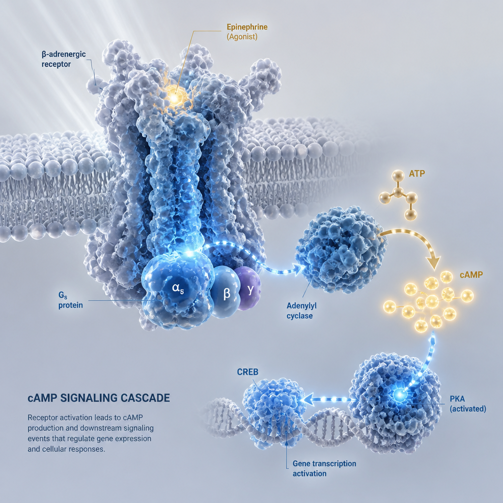
The GLP-1R is a class B G protein-coupled receptor (GPCR). Its primary coupling partner is the Gs alpha subunit. Upon agonist binding, GLP-1R activates adenylate cyclase, which catalyzes the conversion of ATP to cyclic adenosine monophosphate (cAMP). This is the signal that propagates downstream and drives the insulin-secretory and cytoprotective effects associated with GLP-1R activation in pancreatic beta cells.
Downstream of cAMP accumulation, protein kinase A (PKA) and the exchange protein directly activated by cAMP (Epac2) are the main effectors. PKA phosphorylates multiple substrates involved in exocytosis of insulin granules. In parallel, Epac2 facilitates Rap1-GTP exchange, contributing to both secretory and proliferative effects in beta cells. The GLP-1R signaling pathway in pancreatic beta cells acts within seconds of receptor activation, making it highly amenable to time-resolved in vitro assay formats.
Beyond beta cells, GLP-1R expression has been confirmed in hypothalamic neurons, hepatocytes, and cardiac tissue. This matters for in vitro researchers selecting cell systems. CHO-K1 cells stably transfected with human GLP-1R are the most common recombinant model for cAMP-based potency assays. INS-1E cells, a rat insulinoma line, offer an endogenously expressing beta-cell proxy but carry the caveat of rat-origin receptor with known species differences in agonist binding. MIN6 cells provide a mouse beta-cell context. Each model system answers different experimental questions and introduces different interpretive constraints.
Pro tip: When designing a retatrutide GLP-1R cAMP assay in CHO-K1 cells, include a GLP-1(7-36) amide reference standard in every plate to normalize for inter-assay variability. The relative EC50 of retatrutide to your reference standard is more reproducible across laboratories than the absolute cAMP signal values.
Structural Basis of GLP-1R Engagement in Retatrutide
Published cryo-EM structural data on retatrutide bound to GLP-1R, GIPR, and GCGR reveals a combination of conserved peptide-receptor interactions and receptor-specific conformations, particularly in extracellular loop 1 (ECL1), that enable retatrutide to execute agonism at multiple receptors. This is mechanistically significant, not decorative structural biology.
The ECL1 Recognition Problem
Each of the three target receptors requires recognition of ECL1 for productive agonism. The structural work shows that six key peptide positions in retatrutide use amino acids similar to those in native GLP-1 and glucagon at the positions that contact ECL1. This conservation is what enables cross-receptor activity. The middle region of the peptide, by contrast, shows considerable variability across dual and triple agonists, reflecting the tuning required to achieve a desired receptor-potency balance.
Retatrutide's GLP-1R potency is approximately 2.5-fold lower than native GLP-1. This is a deliberate pharmacological trade-off, not a manufacturing limitation. The reduced GLP-1R potency relative to semaglutide or liraglutide means that the glucagon receptor contribution becomes proportionally more important to the total energy expenditure effect. For in vitro researchers, this potency relationship must be factored into dose selection. Do not apply GLP-1 EC50 assumptions to a retatrutide GLP-1R assay and expect matched signal intensity.
Biased Signaling Considerations
Biased agonism, where a ligand preferentially activates one downstream pathway (such as Gs-cAMP) over another (such as beta-arrestin recruitment), is an increasingly important concept in GLP-1R pharmacology. A compound that is biased away from beta-arrestin recruitment tends to show reduced receptor desensitization and internalization, which has implications for sustained signaling in longer in vitro incubations. Whether retatrutide exhibits meaningful GLP-1R biased agonism relative to GLP-1 is an open experimental question that represents a tractable in vitro research opportunity.

In Vitro Research Implications: Cell Models, Assays, and Endpoints
Translating GLP-1R mechanism knowledge into productive in vitro research with retatrutide requires matching the right cell model to the right experimental question. The most common mismatch seen in the literature is applying a recombinant overexpression system when the question is about physiologically relevant receptor density, or vice versa.
Cell Model Selection
For receptor potency characterization and EC50 determination, CHO-K1 cells stably expressing human GLP-1R are the appropriate system. They provide clean, interpretable cAMP data with minimal confounding from endogenous GIPR or GCGR expression. For studying GLP-1R-mediated insulin secretion, INS-1 or MIN6 cells retain more physiological relevance but require careful confirmation of receptor expression levels and species-specific pharmacology differences. For hepatic lipid metabolism questions, primary hepatocyte cultures or HepG2 cells with confirmed GLP-1R expression are used in the published literature on GLP-1 receptor agonist effects on NAFLD-relevant endpoints.
Assay Formats for GLP-1R Activation
The standard approach for confirming GLP-1R functional activity is a cell-based cAMP accumulation assay. Commercially available formats include HTRF (homogeneous time-resolved fluorescence), LANCE Ultra, and HitHunter cAMP assay kits. Each format can be run in 384-well plate configurations, making them compatible with dose-response curve generation across a range of retatrutide concentrations. The key endpoint is EC50 and Emax relative to the GLP-1(7-36) reference standard.
Beyond cAMP, beta-arrestin recruitment assays (PathHunter, PRESTO-Tango) allow assessment of biased agonism. Insulin secretion assays from beta-cell lines provide functional metabolic readouts. Receptor internalization assays using fluorescently tagged GLP-1R constructs add a trafficking dimension. Each of these endpoints requires its own compound concentration optimization and should not be assumed to mirror cAMP assay parameters.
Pro tip: In retatrutide in vitro cAMP assays, always include a GCGR-selective antagonist and a GIPR-selective antagonist as negative controls when working in cell lines that express all three receptors. Without receptor-specific pharmacological blockade, you cannot attribute the observed cAMP response to GLP-1R alone, regardless of what your study design intends to show.
Comparing GLP-1R Engagement Across Single, Dual, and Triple Agonists
Understanding how retatrutide's GLP-1R engagement profile differs from classical GLP-1 monotherapy and dual agonism is essential context for interpreting in vitro data and for communicating results accurately.
| Compound Class | Receptor Targets | GLP-1R Potency and In Vitro Research Considerations |
|---|---|---|
| GLP-1 Monotherapy (e.g., semaglutide, liraglutide) | GLP-1R only | Full GLP-1R selectivity. All cAMP signal attributable to GLP-1R. Provides the cleanest in vitro mechanistic readout for GLP-1R biology. Reference standard class for comparative potency. |
| GLP-1/GIP Dual Agonist (e.g., tirzepatide) | GLP-1R + GIPR | Moderate GLP-1R potency with GIPR co-activation. In vitro requires two-receptor controls. Structural work on tirzepatide has informed understanding of the shared ECL1 interaction motif that retatrutide also uses. |
| GLP-1/GIP/Glucagon Triple Agonist (retatrutide) | GLP-1R + GIPR + GCGR | Approximately 2.5x lower GLP-1R potency than native GLP-1. All three receptor pathways active simultaneously. In vitro demands receptor-specific controls. Glucagon receptor contribution adds energy expenditure signaling absent in mono or dual agonists. |
Compound Handling and Purity Considerations for GLP-1R Research
The pharmacological precision of GLP-1R in vitro research is only as reliable as the compound you start with. This is not a generic quality statement. It is specific to retatrutide for a structural reason: the C20 fatty diacid moiety at lysine-20 makes this a more complex molecule to synthesize and purify than simpler GLP-1 analogs. Impurities in a retatrutide lot are not inert. Truncated or modified peptide sequences can act as partial agonists, antagonists, or silent binders at GLP-1R, silently distorting your EC50 or Emax values.
For mechanistic in vitro GLP-1R research, 99% or higher purity, verified by an independent analytical laboratory, is the minimum acceptable standard. Certificate of Analysis data should include HPLC purity trace, mass confirmation by LC-MS, and water content. Batch traceability, meaning the ability to trace your specific research lot back to a verified synthesis and QC record, allows you to exclude compound quality as a confounding variable when you submit your data for peer review.
Lyophilized format is the practical standard for research-grade retatrutide. It provides long-term stability, allows precise reconstitution at defined molar concentrations, and is compatible with the sub-milligram quantities typical of in vitro assay work. Reconstitution should be done in sterile aqueous solvent with gentle mixing, not vortexing, to avoid aggregation of the fatty diacid moiety. Prepare working aliquots immediately after reconstitution and avoid repeated freeze-thaw cycles, which degrade peptide integrity and introduce assay-to-assay variability.
The mechanistic conclusions drawn from any GLP-1R in vitro study are contingent on the pharmacological identity of the compound used. Purity documentation is not paperwork. It is part of the experimental method.
When sourcing retatrutide for GLP-1R research, the standard you should hold suppliers to includes: independent Canadian laboratory verification (not just in-house QC), documented chain of custody from synthesis to delivery, and lot-specific analytical data available before purchase. Pepura Labs provides lyophilized retatrutide at 99%+ purity, with batch traceability and Canadian laboratory-verified CoA documentation, specifically structured to meet the evidentiary standards that mechanistic in vitro research requires.
Frequently Asked Questions
What does GLP-1R agonism specifically contribute to retatrutide's mechanism compared to its GIPR and glucagon receptor activity?
GLP-1R agonism in retatrutide drives glucose-stimulated insulin potentiation in pancreatic beta cells, hypothalamic appetite suppression, and slowed gastric motility signaling. These are the same downstream effects seen with GLP-1 monotherapy but at approximately 2.5x lower receptor potency in retatrutide. The GIPR component adds incretin co-signaling and modulates GLP-1R-induced nausea pathways. The GCGR component contributes hepatic glucose output regulation and thermogenic energy expenditure signaling that is absent from GLP-1 monotherapy. All three components act simultaneously, making receptor attribution a design challenge in multi-receptor-expressing cell systems.
Why is cAMP the primary in vitro readout for GLP-1R activation rather than another endpoint?
GLP-1R is primarily coupled to the Gs alpha subunit of heterotrimeric G proteins. Gs coupling activates adenylate cyclase, producing cAMP as the direct, proximal second messenger. This makes cAMP accumulation the most direct and most rapidly established functional readout for GLP-1R engagement. Downstream endpoints like insulin secretion, beta-arrestin recruitment, or receptor internalization are valid but add mechanistic steps between receptor activation and measurement, each of which introduces additional biological and technical variables that complicate attribution.
We would love your feedback and any insights you would share with others. What perspective would you add?
Can I use a standard GLP-1 EC50 value as a reference to set my retatrutide in vitro dose range?
Not reliably. Retatrutide's GLP-1R potency is approximately 2.5-fold lower than native GLP-1. If you anchor your dose range to a GLP-1 EC50 and apply the same concentration range to retatrutide expecting equivalent GLP-1R activation, you will underestimate the concentration needed for full GLP-1R efficacy. Always generate a full dose-response curve for retatrutide with a co-run GLP-1(7-36) reference standard and report EC50 values relative to that standard rather than as absolute values compared to historical GLP-1 data.
What cell lines are most appropriate for studying retatrutide's GLP-1R component specifically?
For GLP-1R-selective mechanistic work, CHO-K1 cells stably transfected with human GLP-1R offer the cleanest system because they lack endogenous GIPR and GCGR. This allows you to isolate GLP-1R-mediated cAMP signaling unambiguously. If you require a more physiologically relevant beta-cell context, INS-1 or MIN6 cells provide endogenous GLP-1R expression, but you must add receptor-selective antagonists to block GIPR and GCGR co-activation if you want to attribute results specifically to GLP-1R. Failing to do so makes mechanistic interpretation ambiguous.
How does retatrutide's C20 fatty diacid structure affect in vitro assay design?
The C20 fatty diacid conjugated at lysine-20 enables albumin binding, which is what extends the compound's half-life to approximately six days in biological systems. In vitro, this means retatrutide will partition into albumin present in serum-supplemented media, reducing the free concentration available to bind GLP-1R. Assays run in serum-free or low-serum media will give higher apparent GLP-1R potency than those in full serum conditions. This is a real confound that affects EC50 comparability across labs. Standardize your serum conditions and report them explicitly in your methods.
What purity threshold is acceptable for retatrutide in GLP-1R in vitro research?
99% or higher, verified by an independent analytical laboratory rather than only by the supplying manufacturer. The fatty diacid modification on retatrutide increases the likelihood of synthesis-related impurities including truncated sequences and oxidation products. Any impurity that retains partial GLP-1R binding activity will distort your EC50 or Emax measurements in ways that are invisible without a purity check. Independent CoA documentation with HPLC trace and LC-MS mass confirmation is the minimum acceptable standard for mechanistic GLP-1R research.
Is there published structural data showing how retatrutide engages GLP-1R specifically?
Yes. Cryo-EM structural data has been published characterizing retatrutide bound to GLP-1R, GIPR, and GCGR. The structural work reveals that conserved peptide-receptor interactions at extracellular loop 1 (ECL1) are critical for productive agonism at all three receptors, and that receptor-specific conformational changes at ECL1 distinguish each receptor engagement. The six key amino acid positions in retatrutide that contact ECL1 are conserved relative to native GLP-1 and glucagon sequences, while the middle region of the peptide shows variability tuned to achieve the desired potency balance across receptors.
If you are working with retatrutide in GLP-1R in vitro research, share your assay design challenges or findings in the comments. Specific technical questions about cell model selection, assay format, or compound handling for this compound class are worth discussing in detail.
References
- Structural insights into triple agonism at GLP-1R, GIPR and GCGR manifested by retatrutide (PubMed Central, Cell Discovery 2024)
- GLP-1 receptor trafficking and signaling in pancreatic beta cells (Frontiers in Endocrinology)
- Retatrutide mechanism of action overview including receptor targets and structural properties (Lilly Medical)
- GLP-1 receptor agonist direct modulation of lipid metabolism in hepatic tissue using in vitro NAFLD models (PubMed Central)
- In vivo and in vitro characterization of a long-acting GLP-1 receptor agonist including cAMP assay methodology (PubMed Central)