
Most researchers approaching GIP receptor biology for the first time focus almost entirely on ligand binding affinity and receptor occupancy, and then wonder why their insulin sensitisation readouts are inconsistent. The real story happens downstream: the GIP receptor secondary messenger cascade that translates GIPR activation into measurable metabolic changes is a layered signalling network, not a single switch. If your insulin sensitisation assay is not designed around the specific nodes of that network, you are measuring noise. This guide is for research teams using high-purity peptide compounds to run rigorous in vitro GIP downstream signalling research, and it covers exactly which assay formats map to which biological signals.
Table of Contents
- Quick Takeaways
- GIP Receptor Signalling: The Cascade You Actually Need to Map
- cAMP: The Primary Secondary Messenger in GIPR Activation
- Downstream Effectors: PKA, Epac, and the PI3K-Akt Axis
- Insulin Sensitisation Assay Formats for GIP Research
- Comparison of Secondary Messenger Detection Methods
- Retatrutide and Multi-Receptor Assay Design Considerations
- Compound Quality and Assay Reproducibility
- Frequently Asked Questions
- References
Quick Takeaways
| Key Insight | Explanation |
|---|---|
| GIPR couples primarily through Gs | GIP receptor activation raises intracellular cAMP via Gs coupling, making cAMP the definitive primary secondary messenger for any GIPR insulin sensitisation assay. |
| cAMP branches into two functional arms | Elevated cAMP activates both PKA and Epac2, and these arms have complementary but distinct roles in glucose-stimulated insulin secretion and insulin sensitisation. Assays that miss Epac2 produce incomplete pictures under insulin-resistance conditions. |
| HTRF cAMP assays are the current practical standard | Homogeneous time-resolved fluorescence (HTRF) assays for cAMP quantification offer high sensitivity, plate-based scalability, and compatibility with primary adipocytes, which are a key cell model for GIPR insulin sensitisation research. |
| BRET-based biosensors add temporal resolution | Bioluminescence resonance energy transfer (BRET) assays allow real-time kinetic monitoring of cAMP in live cells, which is valuable when studying receptor desensitisation and the time-course of downstream insulin sensitisation signals. |
| PI3K-Akt phosphorylation is the downstream insulin sensitisation endpoint | After cAMP rises, the PKA and Epac arms converge on PI3K-Akt activation, IRS-1 phosphorylation, and GLUT4 translocation. Phospho-Akt (Ser473) is the most actionable single endpoint for an insulin sensitisation assay. |
| Beta-arrestin recruitment is weak at GIPR | Unlike GLP-1R, GIPR shows minimal beta-arrestin 2 recruitment in most cell systems. Designing arrestin-based assays for GIPR without accounting for this will produce flat dose-response curves and false negatives. |
| Peptide purity directly affects EC50 reliability | Impurities in research-grade GIP or triple-agonist peptides (such as Retatrutide) shift apparent EC50 values, compress dynamic range in cAMP assays, and introduce systematic error into any downstream insulin sensitisation assay. |
GIP Receptor Signalling: The Cascade You Actually Need to Map
The glucose-dependent insulinotropic polypeptide receptor (GIPR) belongs to the secretin subfamily of G protein-coupled receptors (GPCRs). It shares significant sequence homology with GLP-1R, which makes it tempting to run the same assay stack across both receptors, but the signalling phenotypes differ in ways that matter for insulin sensitisation research.
GIPR and GLP-1R are closely related members of the secretin family of GPCRs. When activated, both receptors couple through Gs, producing an intracellular cAMP rise that potentiates glucose-stimulated insulin secretion. The downstream signal from that cAMP elevation is where the biology becomes specific to each receptor, and where assay design decisions have real consequences for data quality.
A common mistake in GIP downstream signalling research is treating GIPR as functionally interchangeable with GLP-1R because they share the Gs/cAMP axis. In practice, the kinetics of cAMP accumulation, the degree of receptor internalisation, and the relative weight of the PKA vs. Epac arms differ between the two receptors, especially in adipocyte and muscle cell models relevant to insulin sensitisation. Running an assay calibrated for GLP-1R on GIPR without re-optimising incubation windows and stimulation concentrations produces systematically compressed cAMP responses.
Pro tip: When setting up a GIPR cAMP assay for the first time, run a full kinetic sweep from 5 to 60 minutes at your target compound concentration before fixing a single endpoint time. GIPR cAMP responses in primary adipocytes peak earlier than in stable overexpression lines, and choosing the wrong time point flattens your dose-response curve before you even test your experimental compound.

cAMP: The Primary Secondary Messenger in GIPR Activation
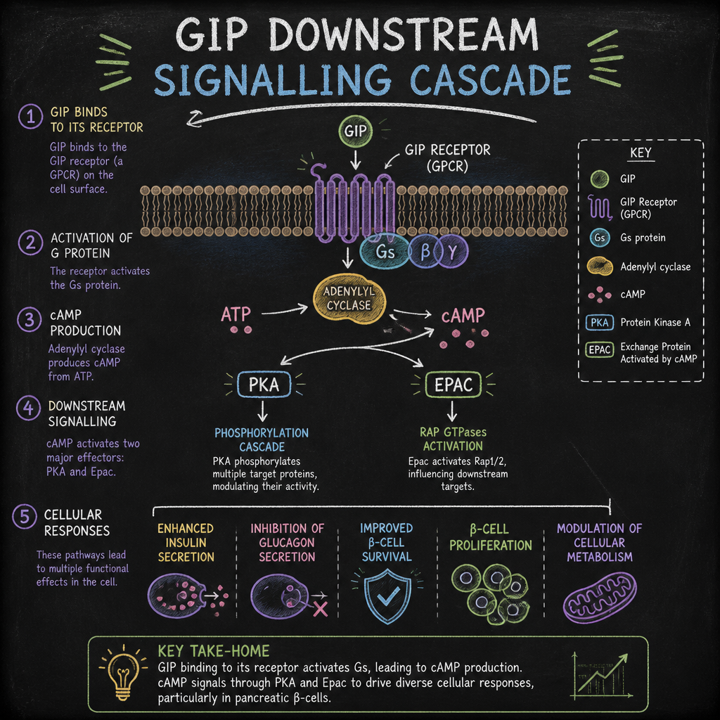
Every serious insulin sensitisation assay built on GIPR biology starts with cAMP. GIP potentiates glucose-stimulated insulin secretion by increasing intracellular cAMP concentration, which is the mechanistic basis for its incretin classification. This cAMP signal is not just a surrogate of receptor activation: it is the functional branch point from which all downstream insulin sensitisation effects originate.
In practical terms, this means your cAMP readout is your assay anchor. Everything else, including PKA substrate phosphorylation, Epac-mediated Rap1 activation, and PI3K-Akt engagement, sits downstream of the cAMP signal and will only be interpretable if you have clean, validated cAMP data first.
Why IBMX Matters More Than Researchers Expect
Phosphodiesterase (PDE) inhibition is not optional in GIPR cAMP assays. cAMP produced after GIPR activation is rapidly degraded by intracellular PDEs, and the GIPR response in primary cells is modest enough that uninhibited degradation can mask real signal. Standard practice uses IBMX (isobutylmethylxanthine) at 0.5 mM in the assay buffer. This concentration is well-established in published GIPR cAMP methodology using HTRF assays in primary mouse and human adipocytes.
The cell model matters too. Stable overexpression lines expressing recombinant GIPR (such as CHO or HEK293 cells) produce higher maximal cAMP responses than primary adipocytes expressing endogenous GIPR, simply due to receptor copy number. If your research objective relates to insulin sensitisation in physiologically relevant tissue, running the assay in a primary adipocyte or muscle cell background, despite the added complexity, gives you data that translates. Endogenous GIPR cell lines, including Neuro-2a neuroblastoma cells as a neuronal model, are used as controls for primary cell comparisons in published GIPR cAMP work.
The cAMP signal produced by GIPR activation is the molecular pivot point between receptor engagement and downstream insulin-sensitising effects. Assays that cannot resolve this signal cleanly cannot resolve anything that follows it.
Downstream Effectors: PKA, Epac, and the PI3K-Akt Axis
Once cAMP rises after GIPR activation, the signal divides into two parallel arms. The first activates protein kinase A (PKA); the second engages Epac2, a cAMP-regulated guanine nucleotide exchange factor. These are not redundant: they drive distinct phosphorylation events with different relevance to insulin sensitisation, and the balance between them shifts under conditions of insulin resistance.
Under normal metabolic conditions, the cAMP-PKA branch dominates and drives glucose-stimulated insulin secretion through phosphorylation of ion channel subunits and insulin granule priming. Under diet-induced insulin resistance, the cAMP-Epac2 arm becomes more prominent. This shift has implications for how you design your peptide research insulin sensitivity assay: if your cell model is insulin-sensitised rather than naive, you need Epac-specific assay components to capture the full signal.
Measuring PKA Activity in GIP Downstream Signalling Research
PKA activity is best measured through phosphorylation of established substrates rather than through the kinase itself. Phospho-CREB (Ser133) is a well-validated nuclear PKA substrate that can be quantified by ELISA or AlphaLISA in a plate-based format compatible with high-throughput GIP downstream signalling research. Cytoplasmic substrates, including phospho-SUR1 in beta cell models, are more mechanistically specific but require more complex cell handling.
A common mistake is using a generic PKA activity assay kit calibrated for adrenergic receptor agonists, which have much larger cAMP amplitudes than GIPR. The sensitivity of off-the-shelf PKA assays is often insufficient to resolve the more modest cAMP rises seen with GIP at endogenous receptor densities. Cell-based PKA FRET biosensors offer better sensitivity in this range.
The PI3K-Akt Axis as an Insulin Sensitisation Endpoint
Downstream of both PKA and Epac2, GIPR activation in adipose and muscle models engages the PI3K-Akt signalling cascade. The IRS-1/PI3K/Akt pathway is the canonical insulin sensitisation route: after PI3K is activated through IRS-1 tyrosine phosphorylation, it generates PIP3, which recruits and activates Akt (phospho-Akt at Ser473 and Thr308). Activated Akt then phosphorylates AS160, facilitating GLUT4 translocation to the sarcolemma and glucose uptake.
For an insulin sensitisation assay anchored to GIPR biology, phospho-Akt (Ser473) is the most actionable single endpoint. It sits at the convergence point of multiple upstream GIP-driven signals, it is quantifiable by standard Western blot, ELISA, or bead-based multiplex, and it has direct mechanistic relevance to insulin sensitisation outcomes. Pairing it with phospho-IRS-1 (Tyr612) gives you the upstream/downstream confirmation needed to distinguish true GIPR-mediated sensitisation from nonspecific Akt activation.
Pro tip: When running phospho-Akt assays downstream of GIPR stimulation, serum-starve your cells for at least 4 hours before treatment. Residual serum growth factors activate Akt constitutively, and this background drowns out the more subtle GIP-mediated phospho-Akt increment, especially in primary adipocytes where GIPR expression is lower than in overexpression systems.
Insulin Sensitisation Assay Formats for GIP Research
The choice of assay format is not just a technical preference. It determines which part of the GIPR signalling cascade you can resolve, at what throughput, and whether the data will hold up to the scrutiny of secondary messenger specificity. Here is what works in practice, and what does not.
HTRF cAMP Assay: The Workhorse
Homogeneous time-resolved fluorescence (HTRF) cAMP assays are the current practical standard for quantifying GIPR-driven cAMP in plate-based formats. The assay principle is competitive: endogenous cAMP competes with a labelled cAMP tracer for an anti-cAMP antibody, and the HTRF signal is inversely proportional to cAMP concentration in the sample. This format has been validated specifically for GIPR work in primary mouse and human adipocytes using serial dilution of agonist concentration, with cells incubated in assay buffer containing BSA and IBMX.
The advantages are real: no cell lysis artefacts if you use the lysis-based protocol correctly, compatibility with 384-well plates for compound screening, and established reference ranges from the published GIPR literature. The limitation is time resolution: you are getting a snapshot at a fixed endpoint, not a kinetic trace.
BRET Biosensors: Real-Time cAMP Kinetics in Live Cells
BRET-based cAMP biosensors allow real-time monitoring of cAMP dynamics in living cells without cell lysis. The BRET signal changes continuously as cAMP rises and falls, making it possible to capture receptor desensitisation kinetics, compare the onset rates of different GIPR agonists, and run time-course experiments that fixed-endpoint HTRF cannot provide.
This format is particularly valuable when studying GIPR desensitisation, which is a relevant variable in any GIP downstream signalling research using repeated or prolonged stimulation, as in multi-receptor agonist protocols. BRET assays require stable cell transfection with the biosensor construct, which adds lead time, but the kinetic data quality justifies the setup cost for mechanistic studies.
FRET Biosensors for Subcellular cAMP Compartmentalisation
FRET-based cAMP sensors, including EPAC-based probes, have been used to measure cAMP precisely in specific intracellular compartments. This matters because cAMP signalling from GPCRs is not uniformly distributed across the cell. Compartmentalised cAMP pools drive different downstream effects, and a cytoplasmic total-cAMP readout misses the spatial dimension of GIPR signalling. EPAC-based FRET probes provide a direct measure of cAMP dynamics with high selectivity and temporal resolution, which has been used to confirm GPCR-mediated cAMP responses in mechanistic signalling studies.
Comparison of Secondary Messenger Detection Methods
| Method | Best Use Case in GIP Research | Key Limitation |
|---|---|---|
| HTRF cAMP Assay | High-throughput compound screening, dose-response curves for GIPR agonists in primary adipocytes or stable lines, quantitative cAMP concentration comparisons across compound batches | Fixed endpoint only; no kinetic resolution; requires IBMX pre-treatment and careful timing to avoid PDE-driven signal collapse |
| BRET cAMP Biosensor | Real-time kinetics of cAMP in live cells; receptor desensitisation studies; comparing onset and offset rates of different GIPR agonists or multi-receptor compounds | Requires stable transfection of biosensor construct; throughput limited compared to HTRF; signal is relative rather than absolute cAMP concentration |
| Phospho-Akt ELISA / Western Blot | Measuring the insulin sensitisation endpoint downstream of cAMP; confirming that GIPR activation translates to the PI3K-Akt axis; validating compound bioactivity in a metabolically relevant readout | Multiple steps removed from receptor activation; sensitive to serum background; does not distinguish GIP-specific Akt activation from off-target kinase activity without receptor-specific controls |
Retatrutide and Multi-Receptor Assay Design Considerations
Retatrutide is a synthetic peptide that acts simultaneously as an agonist at three receptors: the GLP-1 receptor, the GIP receptor (GIPR), and the glucagon receptor (GCGR). This multi-receptor profile makes it an important research tool for studying how simultaneous GIPR and GLP-1R co-activation modulates insulin sensitisation signals compared to single-receptor agonism, but it introduces assay design challenges that do not arise with GIP alone.
When using Retatrutide in a GIP receptor secondary messenger assay, the cAMP signal you detect integrates contributions from all three activated receptors. In cells co-expressing GLP-1R and GIPR (which includes most beta cell models and many adipocyte preparations), the combined cAMP response is larger in amplitude than GIP-only stimulation, and the dose-response curve shape reflects the composite pharmacology. This is not an artefact: it is the biology. But if your objective is to isolate the GIPR-specific contribution to insulin sensitisation, you need receptor-specific antagonist controls or cell lines with single-receptor expression.
Research published on GLP-1R and GIPR heteromer formation using FRET and BRET saturation experiments confirms that these two receptors can form heteromers in cell lines such as HEK-293. Stimulation of these heteromers with GLP-1 increases FRET and BRET ratios, while GIP stimulation decreases them, indicating that the signalling geometry changes when both receptors are present together. Retatrutide studies in co-expression systems should account for this interaction, particularly when interpreting cAMP plateau values or comparing them to mono-agonist benchmarks.
From a practical standpoint, the dosage format of Retatrutide used in in vitro research matters. Lyophilised preparations at defined dosages, such as the 10 mg, 20 mg, 40 mg, 60 mg, and 84 mg formats available from Canadian research suppliers like Pepura Labs, allow researchers to reconstitute to precise molar concentrations with bacteriostatic water, which is critical for generating reproducible concentration-response curves in secondary messenger assays. Any variation in actual peptide content from labelled quantity directly propagates into systematic errors in EC50 determination.
Compound Quality and Assay Reproducibility
This is where many research programmes quietly fail. The downstream insulin sensitisation assay design can be meticulous, the cell model can be appropriate, and the detection technology can be well-suited to GIPR biology, and still the data will be irreproducible if the peptide compound used to stimulate the receptor is not of sufficient purity or does not come with verified lot-specific characterisation.
For GIP downstream signalling research, 99%+ purity is the practical minimum for generating publishable secondary messenger data. Impurities in research-grade GIPR agonist preparations are not inert: they may compete for receptor binding, activate off-target GPCRs, or degrade to products that act as partial agonists or inverse agonists. Any of these outcomes shifts your measured cAMP EC50, distorts your phospho-Akt dose-response curve, and makes inter-lot comparison unreliable.
In Canada, research teams have access to Pepura Labs compounds that are independently verified by Canadian laboratories and supplied with full Chain of Custody documentation, including batch-specific certificates of analysis (COAs). This matters operationally because it allows you to trace any anomalous secondary messenger assay result back to lot-level quality data rather than spending time troubleshooting assay variables that are actually compound variables.
Reconstitution practice also affects assay quality. Lyophilised peptides reconstituted with standard water will degrade in days under refrigeration due to microbial contamination. Using bacteriostatic water for reconstitution and storing aliquots at the correct temperature are basic steps, but they are the difference between consistent EC50 values across a six-week assay campaign and drift that you cannot explain.
Pro tip: Request batch-specific mass spectrometry data alongside your COA for any GIPR agonist compound you use in secondary messenger assays. Purity percentage by HPLC is a necessary but not sufficient quality check: MS confirmation of the correct molecular mass rules out co-eluting impurities at the same retention time that HPLC alone would miss.
Frequently Asked Questions
What is the GIP receptor secondary messenger and why does it matter for insulin sensitisation research?
The primary secondary messenger for the GIP receptor (GIPR) is cyclic AMP (cAMP), produced when activated GIPR couples through the Gs protein to stimulate adenylyl cyclase. This cAMP signal activates PKA and Epac2, which in turn engage the PI3K-Akt pathway central to insulin sensitisation. Without accurately measuring this cAMP signal, and its downstream effects on PKA and Epac substrates, you cannot reliably attribute observed insulin sensitisation outcomes to GIPR activation specifically.
Why does the choice between PKA and Epac readouts matter in a GIP insulin sensitisation assay?
PKA and Epac2 have complementary but distinct roles in GIPR-mediated biology. Under normal conditions, the PKA arm drives the majority of glucose-stimulated insulin secretion enhancement. Under insulin resistance conditions, the Epac2 arm becomes more prominent in controlling that same response. If your cell model is metabolically stressed or insulin-resistant, an assay reading only PKA activity will underestimate total GIPR-driven signalling output. Using both a PKA substrate readout and an Epac-specific assay (such as Rap1-GTP pull-down or an Epac FRET sensor) gives the complete picture.
What cell models are most appropriate for a peptide research insulin sensitivity assay targeting GIPR?
Primary adipocytes (mouse or human) expressing endogenous GIPR are the gold standard for physiological relevance because they reflect native receptor density and lipid metabolism context. For mechanistic and screening work, stable HEK293 or CHO lines overexpressing GIPR offer higher signal amplitude and greater throughput, but data from these lines should be validated in at least one primary cell format before being treated as physiologically predictive. Beta cell lines expressing both GIPR and GLP-1R are appropriate when the research question concerns incretin co-signalling rather than isolated GIPR biology.
Does beta-arrestin recruitment play a useful role in GIPR secondary messenger assay design?
In most cell systems studied to date, GIPR shows very weak or undetectable beta-arrestin 2 (arrestin3) recruitment in response to GIP stimulation. This is in contrast to GLP-1R, which recruits beta-arrestin in a dose-dependent manner. Designing a GIPR assay around beta-arrestin as a primary readout will yield flat curves and false negatives. Arrestin-based assays at GIPR may be useful as negative controls or for studying specific receptor mutants, but they should not be the primary endpoint in a standard GIP downstream signalling research protocol.
How does compound purity affect secondary messenger assay results in GIP research?
Peptide purity affects every quantitative parameter in a secondary messenger assay. Impurities in GIPR agonist preparations can compete for receptor binding, activate other GPCRs present in your cell model, or behave as partial agonists that compress the maximum cAMP response. The practical result is shifted EC50 values, reduced Emax, and irreproducible inter-lot comparisons. For in vitro GIP downstream signalling research, compounds verified at 99%+ purity by HPLC and confirmed by mass spectrometry, supplied with a lot-specific COA, are the minimum standard for data you intend to publish or use for downstream mechanistic conclusions.
What is the correct approach to detecting PI3K-Akt activation downstream of GIPR stimulation in a cell-based insulin sensitisation assay?
Serum-starve cells for a minimum of 4 hours before stimulation to suppress constitutive Akt activity driven by serum growth factors. Stimulate with your GIPR agonist at multiple concentrations spanning at least three orders of magnitude. Measure phospho-Akt at Ser473 and, if resources allow, Thr308 as well, since full Akt activation requires dual phosphorylation. Pair this with phospho-IRS-1 (Tyr612) to confirm upstream pathway engagement. Include a receptor-specific antagonist control and an insulin positive control to bracket your GIPR-driven Akt signal within a known physiological range.
If your research involves Retatrutide or other compounds that target multiple receptors simultaneously alongside GIPR, we would be glad to hear how your assay design has handled the multi-receptor cAMP integration problem. Share your experience or questions in the comments.
References
- GLP-1 and GIP receptor signalling in human beta cells: cAMP production and incretin-driven insulin secretion mechanisms
- GIPR and GLP-1R Gs coupling, cAMP dose-response characterisation, and beta-arrestin recruitment comparison using BRET assays
- HTRF dynamic cAMP assay methodology for GIPR in primary mouse and human adipocytes with endogenous receptor expression
- FRET and BRET analysis of GLP-1R and GIPR heteromerisation and differential cell signalling effects in HEK-293 cells
- Review of incretin peptide hormones including GIP, GLP-1, and glucagon receptor agonist pharmacology and downstream signalling