
The prevailing assumption in GLP-1 appetite suppression research has been that reducing hunger is largely a one-receptor story. Activate the GLP-1 receptor, slow gastric emptying, dampen hunger signaling in the hypothalamus, and the caloric deficit follows. Retatrutide, the investigational triple agonist targeting GLP-1, GIP, and glucagon receptors simultaneously, has forced a significant revision of that model. The appetite-suppressing effects observed in phase 2 trials are not simply "more GLP-1." They emerge from a coordinated, multi-receptor interplay that raises genuinely new questions for researchers studying satiety biology and metabolic peptide pharmacology.
Table of Contents
- Quick Takeaways
- GLP-1 Receptor Signaling: The Core Appetite Mechanism
- How Retatrutide Triple Agonism Reshapes Satiety Research
- Hypothalamic GLP-1 Signaling: What the Evidence Shows
- GIP and Glucagon Receptor Contributions to Appetite Control
- Comparing Appetite Suppression Mechanisms Across Peptide Approaches
- Research Implications: What Retatrutide Studies Tell Us
- Frequently Asked Questions
- References
Quick Takeaways
| Key Insight | Explanation |
|---|---|
| GLP-1 receptor agonism is the foundation of appetite suppression in retatrutide | GLP-1R activation reduces hunger signaling in the hypothalamus, slows gastric emptying, and reduces meal absorption rate, producing a sustained caloric deficit. |
| Hypothalamic GLP-1 signaling is not purely peripheral | GLP-1 is produced centrally by neurons in the nucleus tractus solitarius (NTS), which project directly into the hypothalamic arcuate nucleus, making appetite suppression a brain-level event as much as a gut-level one. |
| GIP receptor co-activation modifies the appetite response in ways GLP-1 alone cannot | Both GIP and GLP-1 receptors are expressed in distinct hypothalamic neuron populations. GIP activation adds a complementary satiety signal that appears to reinforce, rather than duplicate, GLP-1 effects. |
| Glucagon receptor agonism shifts energy balance, not just appetite | GCGR activation increases thermogenesis and resting energy expenditure, meaning retatrutide drives a caloric deficit from two directions: reduced intake and increased expenditure. |
| Phase 2 retatrutide data showed weight reductions of 22.8% and 24.2% at 8 mg and 12 mg doses respectively over 48 weeks | These figures exceed what single or dual agonist approaches have produced in comparable trial windows, suggesting the multi-receptor satiety architecture is genuinely additive. |
| Peptide appetite research now requires multi-circuit models | Vagal afferents, hypothalamic nuclei, brainstem projections, and peripheral incretin signals all interact. Retatrutide studies make it clear that isolating any single pathway understates the biology. |
| Research-grade compound purity matters for mechanistic studies | Off-target receptor activation from impure peptide preparations can confound in vitro satiety signaling data significantly. 99%+ purity with batch traceability is not optional in serious mechanistic research. |
GLP-1 Receptor Signaling: The Core Appetite Mechanism
Before retatrutide can be understood, the foundational biology of GLP-1 appetite suppression research must be clear. Glucagon-like peptide-1 is released from intestinal L-cells in response to nutrient ingestion. It then acts on GLP-1 receptors distributed across multiple tissues, including the gastrointestinal tract, pancreas, vagal nerve terminals, and the brain. The appetite-suppressing effect is not a single event but a cascade operating across at least three anatomical sites simultaneously.
At the gut level, GLP-1 receptor activation slows gastric emptying, extending the mechanical and chemical sensation of fullness after a meal. At the vagal level, GLP-1 signals travel up the afferent vagus nerve to the nucleus tractus solitarius in the brainstem. From the NTS, signals are relayed into hypothalamic circuits that govern hunger and satiety. This is where the central appetite-regulating machinery sits.
The Peripheral vs. Central Distinction in GLP-1 Research
A persistent question in peptide appetite research is whether the clinically observed reduction in food intake comes primarily from peripheral gut signaling or from direct central nervous system effects. The evidence now points firmly toward a dual mechanism. GLP-1 produced peripherally crosses into the brain through circumventricular organs and also acts on vagal afferents that carry the signal centrally, while centrally produced GLP-1 from NTS neurons acts directly on hypothalamic circuits without any peripheral routing at all.
This distinction matters for in vitro research design. Assay systems that only model gut-level receptor binding are capturing roughly half the biological picture. Understanding how GLP-1R agonists operate centrally requires models that account for hypothalamic circuit activation, not just incretin receptor binding assays.
Pro tip: When designing in vitro studies of GLP-1 receptor binding and downstream signaling, use cell lines or primary cultures that express endogenous receptor densities comparable to hypothalamic tissue rather than overexpression systems, which can produce artifactually exaggerated Emax values that do not translate to in vivo readouts.

How Retatrutide Triple Agonism Reshapes Satiety Research
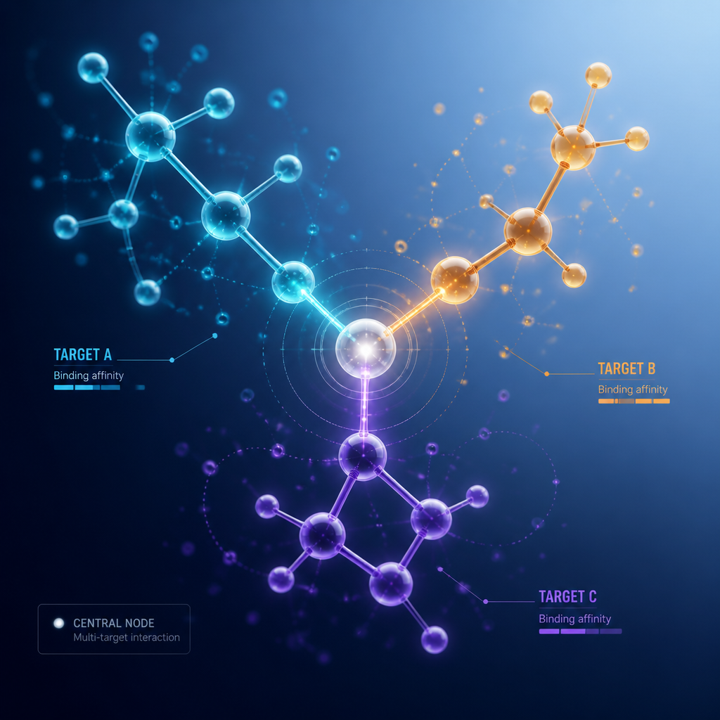
Retatrutide (LY3437943) is a simultaneous agonist of the GLP-1, GIP, and glucagon receptors. In the published phase 2 obesity trial appearing in the New England Journal of Medicine (2023), participants receiving 8 mg of retatrutide achieved a mean weight reduction of approximately 22.8% over 48 weeks, while those on 12 mg achieved approximately 24.2%. One hundred percent of participants in both the 8 mg and 12 mg groups achieved a weight reduction of 5% or more. These are outcomes that outpace the weight-loss magnitude seen with single-agonist GLP-1 therapies in comparable timeframes.
The research question that follows is mechanistic: how much of this effect is appetitive reduction versus changes in energy expenditure? The answer is, deliberately, both. Retatrutide's receptor profile was engineered to produce additive effects across appetite suppression, gastric emptying, insulin secretion, and resting metabolic rate. For researchers studying Retatrutide satiety mechanism, this layered architecture is both the scientific value of the compound and its analytical challenge.
Why the Triple Agonist Design Changes What Researchers Measure
When studying a GLP-1 mono-agonist, changes in food intake can be attributed with reasonable confidence to GLP-1R-mediated pathways. With retatrutide, the observed appetitive suppression reflects simultaneous contributions from all three receptor axes. Parsing the relative contribution of each receptor to the total appetite-suppressive effect requires careful experimental design, including receptor-selective antagonist co-treatment and targeted gene silencing approaches.
In vitro studies using retatrutide can therefore serve a different purpose than efficacy confirmation. They can be used to probe which receptor contributes what proportion of the downstream cAMP generation, which signaling cascade dominates at different ligand concentrations, and how receptor desensitization kinetics differ across the three targets. These are the kinds of questions that high-purity, research-grade retatrutide preparations make tractable.
The complexity of retatrutide's appetite-suppressive activity is a feature for researchers, not a problem to be controlled away. Each receptor axis represents a separable line of inquiry into satiety circuit biology.
Hypothalamic GLP-1 Signaling: What the Evidence Shows
Hypothalamic GLP-1 signaling is now understood to be more anatomically specific than early research suggested. GLP-1 receptor expression is not uniform across the hypothalamus. It is concentrated in specific nuclei, particularly the arcuate nucleus (ARC), the paraventricular nucleus (PVN), and the dorsomedial hypothalamus. The arcuate nucleus is especially relevant to appetite research because it contains two critically opposing neuron populations: pro-opiomelanocortin (POMC) neurons, which suppress appetite, and agouti-related protein (AgRP) neurons, which stimulate it.
Research published in Cell and Bioscience confirmed that GLP-1R neurons are broadly distributed in the brain with particular density in the hypothalamus, including the arcuate nucleus, a region known to regulate energy homeostasis and feeding behavior, which receives dense projections from NTS neurons expressing the proglucagon gene (NTSGcg neurons). This NTS-to-ARC projection pathway is a core anatomical substrate for GLP-1 mediated appetite suppression.
The POMC Neuron Connection in Satiety Research
GLP-1R activation in the arcuate nucleus promotes POMC neuron activity. POMC-derived peptides, including alpha-melanocyte-stimulating hormone, act on melanocortin 4 receptors in the paraventricular nucleus to reduce food intake. This is a well-characterized satiety circuit. What retatrutide studies add is evidence that GIP receptor activation also engages satiety neurons in the hypothalamus through its own receptor population, distinct from but overlapping with GLP-1R-expressing neurons.
The practical implication for peptide appetite research is that the full appetitive phenotype of a triple agonist like retatrutide cannot be captured by measuring output from a single neuron type. Researchers need to think in terms of circuit-level interactions, not single-receptor endpoint assays.
Pro tip: When sourcing retatrutide for hypothalamic cell culture experiments, verify that the supplier provides chain of custody documentation and independent lab verification, not just internal purity claims. Batch-to-batch consistency in a lyophilized peptide directly affects the reproducibility of any dose-response curve you generate against hypothalamic receptor preparations.

GIP and Glucagon Receptor Contributions to Appetite Control
GIP (glucose-dependent insulinotropic polypeptide) has historically been underestimated in appetite research. It was well-characterized as an incretin with effects on pancreatic beta-cell insulin secretion, but its central satiety role was not fully appreciated until research into GIP/GLP-1 co-agonists began revealing outcomes that exceeded what GLP-1 mono-agonism could explain. Research published in Frontiers in Endocrinology clarified that both GIP and GLP-1 activate their respective receptors in distinct neurons within the central nervous system, specifically the hypothalamus, to increase the sense of satiety.
The GIP receptor's presence in hypothalamic neurons means GIP agonism produces genuine central appetite suppression, not merely peripheral metabolic effects. In retatrutide, because it functions as at least a partial agonist at the GIP receptor, this central satiety contribution is present alongside the more established GLP-1R-mediated hunger reduction.
Glucagon Receptor Agonism: Energy Expenditure as Appetite's Complement
The glucagon receptor axis in retatrutide operates differently from GLP-1 and GIP in terms of its appetite relevance. GCGR agonism does not directly suppress hunger signaling in the same way GLP-1R activation does. Instead, it raises resting energy expenditure through thermogenesis and promotes lipid mobilization. The net effect is that the caloric deficit required for weight loss is achieved through a combination of reduced caloric intake (GLP-1R and GIPR mediated) and increased caloric expenditure (GCGR mediated). In research terms, these are mechanistically distinct and separable contributions to the overall metabolic outcome.
Retatrutide's relatively lower activation potency at the glucagon receptor compared to its GIPR effect appears intentional. It provides a metabolic rate contribution without the hepatic glucose production stimulation that higher-potency glucagon agonism would produce, a design trade-off that researchers studying GCGR physiology can probe experimentally using selective antagonists.
Comparing Appetite Suppression Mechanisms Across Peptide Approaches
| Approach | Primary Appetite Mechanism | Research Relevance for Satiety Studies |
|---|---|---|
| GLP-1 Mono-Agonism (e.g., semaglutide-class) | GLP-1R activation in hypothalamus and brainstem; gastric emptying delay; vagal afferent signaling. Single receptor axis, well-characterized downstream cAMP and beta-arrestin pathways. | Cleanest mechanistic model for isolating GLP-1R-specific satiety biology. Limited for studying receptor cross-talk or multi-axis satiety circuits. Mature literature base. |
| GLP-1/GIP Dual Agonism (e.g., tirzepatide-class) | Combined GLP-1R and GIPR activation in hypothalamic neurons; additive central satiety signaling. GIP contribution increasingly shown to enhance, not merely parallel, GLP-1 effects on food intake. | Allows direct study of GIP-GLP-1 receptor cross-talk in satiety circuits. Useful for dissecting incretin-axis contributions to appetite reduction. Phase 3 data well-documented. |
| GLP-1/GIP/Glucagon Triple Agonism (Retatrutide) | Full triple-receptor activation: GLP-1R for primary hunger suppression, GIPR for central satiety augmentation, GCGR for thermogenesis and energy expenditure elevation. Most complex mechanistic profile. | Highest research complexity. Enables study of GCGR-GIPR-GLP-1R crosstalk. Ideal for multi-receptor satiety models. Phase 2 data published. Investigational status means no approved clinical use yet. |
The comparison above clarifies why retatrutide represents a step-change in what researchers can ask about appetite biology. Each successive generation of peptide complexity adds an experimental variable, and a research question. The mono-agonist is a scalpel; the triple agonist is the whole instrument panel.
Research Implications: What Retatrutide Studies Tell Us
The most important insight from retatrutide research for the broader field of GLP-1 appetite suppression research is that the ceiling on appetite suppression from single-receptor approaches was not a biological ceiling. It was an architectural one. By adding receptor axes that independently contribute to central satiety signaling and peripheral energy balance, it becomes possible to produce outcomes that compound rather than merely combine.
For laboratory researchers, this creates a specific set of in vitro priorities. Receptor selectivity profiling using high-purity retatrutide preparations allows investigators to map which of the three receptor axes dominates at each concentration range. Downstream signaling divergence studies, comparing cAMP kinetics, beta-arrestin recruitment, and receptor internalization rates across GLP-1R, GIPR, and GCGR in hypothalamic cell models, can begin to answer which signaling event is most causally responsible for specific behavioral outcomes.
The Purity Standard That Research Requires
In practice, the reproducibility of any in vitro satiety signaling study using retatrutide depends directly on peptide purity. At sub-micromolar concentrations used to model physiological receptor engagement, even a 3% impurity load can introduce receptor cross-reactivity that obscures which receptor is driving the observed signal. This is not a theoretical concern. It is the most common source of contradictory retatrutide dose-response data across different laboratory groups.
Research-grade retatrutide with verified 99%+ purity, backed by independent Canadian laboratory verification and full chain of custody documentation, removes this confound before it contaminates your results. Pepura Labs provides exactly this standard for Canadian research institutions, with lyophilized retatrutide peptide products that carry batch traceability and third-party analytical verification, not just internal quality claims.
A common mistake in peptide appetite research is treating supplier purity certificates as interchangeable. They are not. A self-reported purity figure from a supplier without independent verification is not the same as a purity figure validated by an external analytical laboratory with documented methodology. For mechanistic satiety research, the difference is consequential.
Gastric Emptying Rate and Its Role in Satiety Measurement
One of the most practically important variables in appetite suppression research is gastric emptying rate, because it determines how long a meal remains a satiety stimulus in the gastrointestinal tract. GLP-1R agonism consistently reduces gastric emptying rate, extending postprandial satiety signaling. In triple agonist research, the question is whether GIPR or GCGR co-activation modifies this effect, and if so, whether additively or independently.
Current data from retatrutide studies suggest that the primary gastric emptying effect is GLP-1R mediated, with GIP contributing a separate central satiety signal rather than further slowing gut transit. This means researchers studying gastric motility endpoints in retatrutide preparations need to design their models to distinguish these contributions, using motility assays in parallel with central receptor binding studies rather than treating them as redundant readouts.
Frequently Asked Questions
What is the primary mechanism by which GLP-1 receptor agonism suppresses appetite?
GLP-1 receptor activation suppresses appetite through two parallel mechanisms: peripherally, it slows gastric emptying and reduces the rate of nutrient absorption, prolonging the mechanical fullness signal after meals; centrally, it activates GLP-1R-expressing neurons in the hypothalamic arcuate and paraventricular nuclei, promoting pro-opiomelanocortin neuron activity and reducing the drive to eat. Both mechanisms contribute to the caloric deficit observed with GLP-1R agonist treatment in research subjects.
How does retatrutide differ mechanistically from semaglutide-class GLP-1 mono-agonists in satiety research?
Retatrutide engages three receptor axes simultaneously: GLP-1R, GIPR, and GCGR. Semaglutide-class compounds act almost exclusively on GLP-1R. In satiety terms, retatrutide adds a GIPR-mediated central satiety contribution and a GCGR-mediated increase in resting energy expenditure that semaglutide does not provide. The result is that the caloric deficit in retatrutide studies comes from both reduced intake and elevated expenditure, whereas mono-agonists primarily reduce intake.
Why is hypothalamic GLP-1 signaling considered a central mechanism rather than just a gut hormone effect?
GLP-1 is produced both peripherally (in intestinal L-cells) and centrally (in neurons of the nucleus tractus solitarius in the brainstem). These centrally produced GLP-1 neurons project directly into the hypothalamic arcuate nucleus, which governs energy homeostasis and feeding behavior. This means GLP-1R agonism in the hypothalamus can occur independently of any gut-derived signal, making hypothalamic GLP-1 signaling a bona fide central appetite regulation mechanism rather than simply a relay of peripheral satiety hormones.
What does the GIP receptor contribute to appetite suppression in retatrutide research?
GIP receptor activation contributes to appetite suppression by engaging distinct hypothalamic neurons that express GIPR. Research indicates both GIP and GLP-1 activate their respective receptors in separate but complementary neuron populations within the central nervous system, particularly the hypothalamus, to increase satiety. This means the GIPR contribution in retatrutide is not redundant with GLP-1R signaling but adds a second, parallel satiety signal operating through a distinct neuronal population.
What purity standard should researchers require for retatrutide peptide used in in vitro appetite mechanism studies?
For mechanistic in vitro studies of satiety signaling, retatrutide should meet a minimum of 99% purity as verified by an independent analytical laboratory, not solely by the supplier. Batch traceability and chain of custody documentation are required to ensure that dose-response data from one experiment can be meaningfully compared to another using material from a different preparation lot. Suppliers who provide only internal purity certification without third-party verification introduce a reproducibility risk that is particularly problematic in multi-receptor satiety mechanism research, where impurities can activate off-target receptors and obscure which receptor axis is driving the observed signal.
Are the appetite-suppressing effects of retatrutide purely dose-dependent?
Phase 2 data shows a clear dose-response relationship in weight outcomes, with higher doses producing greater weight reduction over 48 weeks. However, in mechanistic research, the dose-response relationship is more nuanced. At lower concentrations, GLP-1R occupancy dominates and the satiety signal profile resembles single agonist behavior. At higher concentrations, GIPR and GCGR contributions become proportionally more significant. For researchers studying the relative receptor contributions to appetite suppression, choosing the correct concentration range is essential to avoid conflating single-receptor with multi-receptor effects.
If you work with GLP-1 receptor agonists or triple agonist peptides in your research program, we'd value hearing which experimental models you've found most informative for separating central versus peripheral satiety mechanisms.
We would love your feedback and any insights you would share with others. What perspective would you add?
References
- Phase 2 randomized trial of retatrutide for obesity published in the New England Journal of Medicine, reporting 48-week weight reduction outcomes across dose groups
- Cell and Bioscience research article on GLP-1R neuron distribution in the brain, hypothalamus, and arcuate nucleus with NTS projections
- PubMed Central review of GIP and GLP-1 signaling pathways, receptor distribution, and effects on central nervous system satiety neurons
- Diabetes, Obesity and Metabolism journal study on self-reported appetite, dietary restraint, and disinhibition in adults with type 2 diabetes treated with retatrutide versus placebo
- PubMed Central study on central GLP-1 produced in the nucleus tractus solitarius and its role in regulating feeding behavior and energy balance through hypothalamic networks