
Most failed in vitro Retatrutide assay runs come down to one of three problems: poor peptide reconstitution, the wrong cell model for the receptor you are actually targeting, or a cAMP readout that saturates before you capture the full concentration-response curve. Retatrutide (LY3437943) is a triple agonist at the GLP-1 receptor (GLP-1R), the glucose-dependent insulinotropic polypeptide receptor (GIPR), and the glucagon receptor (GCGR), and its differential potency across those three targets means that a single assay design rarely works cleanly for all three. This guide lays out the practical decisions you need to make before you pipette anything, and flags the specific failure modes that waste reagents and delay results.
Table of Contents
- Quick Takeaways
- Understanding Retatrutide Pharmacology Before You Design Your Assay
- Peptide Reconstitution: The Step That Breaks More Assays Than Any Other
- Cell Model Selection for Triple-Receptor Coverage
- Assay Readout Comparison: cAMP, Beta-Arrestin, and Luciferase Reporter
- Assay Buffer Composition and IBMX Concentration
- Concentration-Response Curve Design and EC50 Determination
- Quality Control: Why Purity and Documentation Matter as Much as Protocol
- Frequently Asked Questions
- References
Quick Takeaways
| Key Insight | Explanation |
|---|---|
| Retatrutide has differential receptor potency | It is approximately 8.9-fold more potent at GIPR than endogenous GIP, but less potent than native ligands at GCGR and GLP-1R. Design your concentration range accordingly for each receptor. |
| Lyophilized powder is the stable starting point | Lyophilized Retatrutide is highly stable during storage. Reconstituted solution stored at 2-8°C is workable for months; frozen reconstituted peptide degrades faster, losing a significant fraction of mass within weeks. |
| Reconstitute with bacteriostatic water, not ultrapure water | Bacteriostatic water significantly extends the usable life of the reconstituted solution compared to ultrapure water. Add it slowly along the vial wall and swirl gently. |
| Use low-bind tubes for working dilutions | At picomolar to low nanomolar concentrations, Retatrutide peptide adsorbs readily onto standard polypropylene. Low-bind plasticware prevents concentration errors that invalidate EC50 data. |
| IBMX is non-negotiable for cAMP assays | Phosphodiesterase inhibition with IBMX (typically 2 mM in assay buffer) amplifies and stabilizes the cAMP signal. Without it, GLP-1R and GCGR responses are severely underestimated. |
| Match cell line to receptor subtype precisely | HEK-293 and CHO cell lines stably expressing individual human receptors are the validated workhorse for Retatrutide functional assays. Transient transfection introduces receptor expression variability that makes EC50 comparisons unreliable across experiments. |
| Purity certification is not optional for quantitative work | Batch-to-batch purity differences in the peptide stock directly shift EC50 values. Working with independently verified, 99%+ purity material and full chain of custody documentation is the only defensible baseline for publishable data. |
Understanding Retatrutide Pharmacology Before You Design Your Assay
Retatrutide is a single peptide conjugated to a fatty diacid moiety that confers prolonged half-life through albumin binding. Its molecular weight is 4,731.33 g/mol. What makes it fundamentally different from dual agonists like tirzepatide is that it simultaneously activates three distinct class B GPCRs, each with a different signaling amplitude and a different EC50 window.
Compared to endogenous hormones, Retatrutide is roughly 8.9-fold more potent at GIPR than native GIP, while being less potent at GCGR and GLP-1R relative to their native ligands. This asymmetry is not a detail to note and move on from. It means your concentration-response curve must span at least three to four orders of magnitude if you intend to characterize activity at all three receptors in a single experimental series. Labs that assume a narrow picomolar window for all three targets routinely miss the GCGR response entirely.
The downstream signaling through all three receptors proceeds primarily through Gs-coupled cAMP accumulation, which is why functional cAMP assays are the standard first-pass readout. Beta-arrestin recruitment is a secondary pathway worth investigating if your research question involves biased agonism or receptor desensitization, but it requires a separate assay format and adds considerable experimental complexity.
Pro tip: Before setting up a full concentration-response experiment, run a single-point pilot at 10 nM Retatrutide across all three receptor cell lines. If the fold-change over vehicle is less than 3-fold at that concentration for any receptor, troubleshoot your cell passage number, receptor expression level, and IBMX concentration before committing to an eight-point curve.

Peptide Reconstitution: The Step That Breaks More Assays Than Any Other
Lyophilized Retatrutide is a white powder that is visually unremarkable. The chemistry underneath that appearance is not. At high purity (99% and above), the peptide is largely intact as supplied, but reconstitution errors introduce degradation, aggregation, or concentration inaccuracy that no amount of careful assay execution downstream can fix.
Choosing Your Reconstitution Solvent
Bacteriostatic water is the correct choice for routine reconstitution. It extends the usable life of your reconstituted stock substantially compared to ultrapure or sterile water alone. If your assay buffer is not compatible with the benzyl alcohol preservative, prepare a concentrated stock in bacteriostatic water and dilute into your assay buffer immediately before use, keeping the benzyl alcohol contribution negligible.
For making highly concentrated stocks intended for serial dilution into aqueous buffers, a small percentage of DMSO (typically 1% or less in the final assay well) is sometimes used to aid initial dissolution of the fatty acid-conjugated peptide. If you use DMSO, include a matched DMSO vehicle control at the same final concentration in every experiment. More than 1% DMSO in the assay well will affect GPCR cell signaling results.
Mechanical Handling During Reconstitution
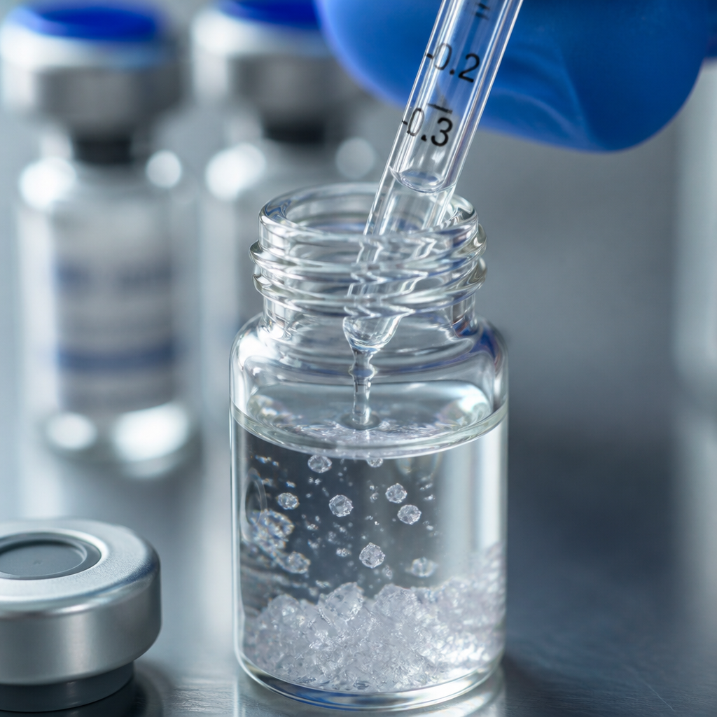
Add your solvent slowly along the inner wall of the vial. Do not pipette directly onto the lyophilized cake. Gently swirl to dissolve. Do not vortex. Do not sonicate. Vigorous mechanical agitation promotes peptide aggregation, which reduces the effective concentration in your working solutions and produces inconsistent results between aliquots drawn from the same vial.
If crystals are visible after initial reconstitution, allow the vial to reach room temperature and swirl gently to redissolve before proceeding.
Storage After Reconstitution
Store reconstituted Retatrutide at 2-8°C, not frozen. Published stability data show that freezing reconstituted peptide solution causes meaningful mass loss (greater than 10% over 45 days), whereas refrigerated reconstituted solution shows single-digit percent mass decline over 60 to 90 days. Prepare aliquots sized for single-use to avoid repeated freeze-thaw cycles, and keep the lyophilized master stock at -20°C until you need it.
Pro tip: Label every aliquot with the date of reconstitution, the lot number, and the concentration. When you get anomalous EC50 data, the first thing to audit is whether you used an aliquot that was beyond its reliable window or one from a different batch. Chain of custody documentation from your peptide supplier makes this audit fast and conclusive.
Cell Model Selection for Triple-Receptor Coverage
The cell model decision is the highest-stakes design choice in a Retatrutide cell assay protocol. You are working with a compound that acts on three different GPCRs with meaningfully different potency at each. Any cell line that endogenously expresses two or more of these receptors will produce mixed signals that are impossible to deconvolute cleanly.
HEK-293 Cells Stably Expressing a Single Human Receptor
HEK-293 cells stably transfected to express a single human receptor (GLP-1R, GIPR, or GCGR) are the established standard for Retatrutide functional agonism assays. The parent HEK-293 line has low endogenous expression of these particular receptors, which gives a clean background. Stable expression provides consistent receptor density from passage to passage, which is essential for comparing EC50 values across experiments or across compound batches.
Culture HEK-293 receptor-expressing lines in DMEM supplemented with 10% FBS. Grow to near confluency in T175 flasks. Detach with accutase rather than trypsin to preserve receptor integrity on the cell surface. Wash with PBS without calcium or magnesium after detachment, then resuspend in assay buffer.
CHO Cell Lines as an Alternative
CHO cells stably expressing individual human receptors are a validated alternative to HEK-293 for cAMP-based functional assays. Their lower background cAMP activity can improve signal-to-noise in some assay formats. For beta-arrestin translocation studies, engineered CHO-K1 PathHunter lines (which express receptor fused to complementary beta-galactosidase fragments) are a practical option for labs that want to characterize biased signaling without building a custom cell line.
What to Avoid
Transient transfection is not suitable for quantitative potency determination. Receptor expression level varies by transfection efficiency and time post-transfection, which shifts apparent EC50 values in ways that are not reproducible. Primary pancreatic beta cells or intestinal organoids that endogenously co-express GLP-1R and GIPR can be useful for physiological studies, but they are not appropriate for isolating individual receptor contributions to Retatrutide's pharmacology.
Assay Readout Comparison: cAMP, Beta-Arrestin, and Luciferase Reporter
Three assay formats dominate the published Retatrutide in vitro literature. Each captures a different aspect of receptor pharmacology and comes with different demands on equipment, throughput, and interpretation.
| Assay Format | Primary Readout | Best Fit for Retatrutide Research |
|---|---|---|
| HTRF cAMP (e.g., Cisbio HTRF kit) | Intracellular cAMP accumulation via fluorescence ratio at 665/620 nm | First-line potency determination (EC50) at each individual receptor. High-throughput compatible, homogeneous format, no wash steps. Industry standard for GLP-1R, GIPR, GCGR agonism profiling. |
| LANCE cAMP (PerkinElmer) | cAMP via time-resolved FRET, read on a TR-FRET-capable plate reader | Alternative to HTRF when Cisbio reagents are unavailable. Comparable sensitivity and dynamic range. Requires specific plate reader capability. Also validated for GLP-1R and GCGR functional assays in the published literature. |
| CRE-Luciferase Reporter / PathHunter Beta-Arrestin | Luminescence via luciferase substrate conversion (CRE-luc) or beta-galactosidase complementation (PathHunter) | Useful for biased agonism characterization and receptor internalization studies. Not interchangeable with cAMP EC50 data. PathHunter requires a specifically engineered cell line; CRE-luciferase requires stable co-transfection with receptor and reporter construct. |
For most labs starting a peptide in vitro research setup with Retatrutide, the HTRF cAMP format is the right starting point. It is homogeneous (no wash), runs in 96- or 384-well format, and produces EC50 data directly comparable to published pharmacology benchmarks. Move to beta-arrestin assays only once you have clean cAMP data establishing your baseline receptor responsiveness.
A compound's EC50 is only as reliable as the cell passage number, the receptor expression level, and the peptide concentration you actually delivered to the well. Control each of these independently before drawing potency conclusions.
Assay Buffer Composition and IBMX Concentration
The assay buffer for Retatrutide cAMP experiments is not negotiable on its core components: 1x HBSS, 20 mM HEPES, 0.1% BSA, and 2 mM IBMX. Each component serves a specific function, and substitutions have consequences.
HBSS (Hanks' Balanced Salt Solution) provides the ionic environment for cell viability during the assay incubation without stimulating intracellular signaling itself. HEPES (20 mM) maintains pH stability independently of CO2, which matters because cAMP assays are run at room temperature outside the incubator. The 0.1% BSA serves as a carrier protein that reduces peptide adsorption to plastic surfaces and stabilizes Retatrutide at low concentrations. Do not omit the BSA if you are working below 1 nM peptide concentrations.
IBMX (3-isobutyl-1-methylxanthine) is a non-selective phosphodiesterase inhibitor. At 2 mM, it prevents intracellular cAMP from being rapidly degraded after receptor activation, which substantially amplifies the detectable signal window. Without IBMX, the cAMP peak is transient and low-amplitude, which compresses the dynamic range of your assay and makes it difficult to distinguish low-potency responses from vehicle baseline. This is especially relevant for GCGR, where Retatrutide's intrinsic potency relative to its native ligand is lower.
Cell seeding density is 400,000 cells/mL in assay buffer, dispensed as 25 µL aliquots into 96-well plates. Compound in assay buffer (25 µL) is added to the wells, followed by incubation for 30 minutes at room temperature. Keep the incubation time consistent across wells. Shorter or longer incubations shift apparent EC50 values systematically.
Pro tip: Always prepare your assay buffer fresh on the day of the experiment. IBMX stock solutions in DMSO (stored at -20°C) are stable, but diluted IBMX in aqueous buffer loses activity over time. Use low-bind 96-well plates, not standard cell-culture-treated plates, for the final assay dispensing step.
Concentration-Response Curve Design and EC50 Determination
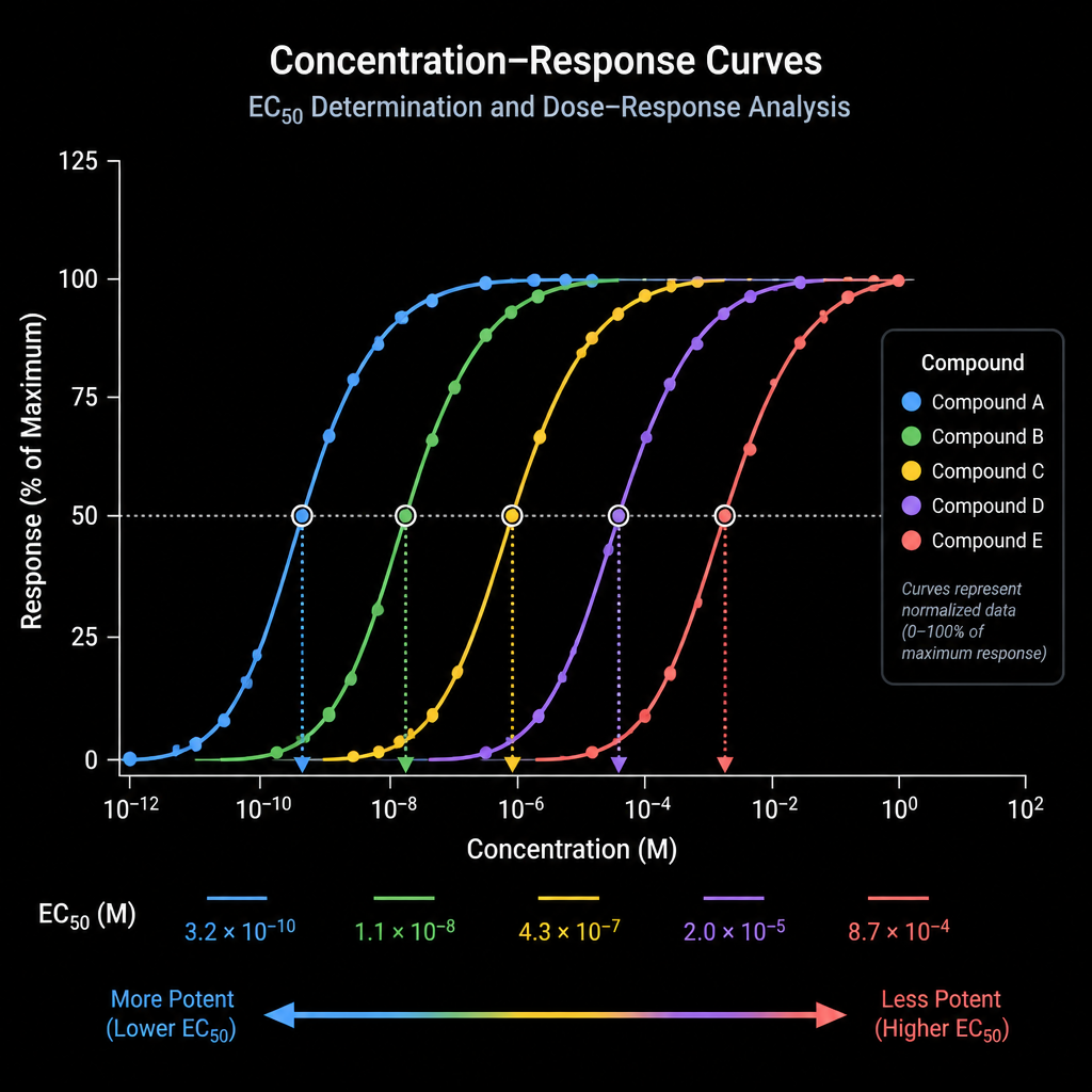
A well-designed concentration-response curve for Retatrutide starts at least two log units below the expected EC50 and extends at least two log units above it. For GIPR, where Retatrutide is highly potent, this means your curve may need to start in the sub-picomolar range. For GCGR, where Retatrutide is less potent relative to native glucagon, your upper concentration may need to extend into the mid-nanomolar range to capture the plateau of the dose-response relationship.
Use an eight- to ten-point curve with half-log or one-log spacing. Serial dilutions should be performed in low-bind microtubes using assay buffer with BSA (not neat water or DMSO stock) as the diluent once you are below 1 µM. This is where a common mistake occurs: labs dilute from a DMSO stock directly into the final assay well without an intermediate dilution in buffered BSA solution, which causes peptide aggregation and concentration errors at the low end of the curve.
Include the following controls in every experiment: vehicle alone (matched DMSO or bacteriostatic water concentration), a maximum stimulation reference using a saturating concentration of the endogenous ligand for each receptor (native GLP-1, GIP, or glucagon), and a known-EC50 reference compound if your lab has one validated from a previous run. These controls let you confirm that the cells are responding normally and that your assay window is consistent before you interpret the Retatrutide curve.
EC50 is calculated by fitting the data to a four-parameter logistic (4PL) curve. Constrain the bottom parameter to the vehicle baseline and confirm that the top plateau reaches a comparable level to the endogenous ligand maximum. If the Retatrutide Emax is substantially lower than the endogenous ligand Emax, investigate whether your top concentration is causing peptide precipitation or receptor desensitization rather than assuming partial agonism without evidence.
Quality Control: Why Purity and Documentation Matter as Much as Protocol
A technically perfect assay protocol produces unreliable data if the peptide stock is impure or undocumented. This is not a theoretical concern. Peptide synthesis byproducts, incomplete deprotection fragments, and oxidized methionine-containing variants all have the potential to occupy the same receptor binding site with different intrinsic efficacies. A batch at 90% purity is not equivalent to a 99%+ purity batch in a quantitative receptor assay.
For Retatrutide specifically, independent verification by a third-party analytical laboratory (HPLC purity, mass confirmation, and ideally amino acid analysis or NMR for identity confirmation) is the standard that serious research programs require. Certificates of Analysis generated by the supplier without independent verification are not equivalent to independently verified batch reports.
Chain of custody documentation matters for a different reason: it allows you to trace any anomalous result back to a specific batch, a specific reconstitution event, or a specific storage period. When your EC50 shifts 5-fold between two experimental series, the first question is whether the peptide stocks were from the same verified batch. Without documentation, that question is unanswerable.
Pepura Labs supplies Retatrutide as lyophilized, independently verified peptide with full chain of custody documentation. Each batch is verified by Canadian analytical laboratories, with HPLC purity at 99% or above confirmed before dispatch. For research programs that need to compare results across time points or across collaborating sites, this level of documentation is not a premium feature. It is the floor of what defensible data requires.
Pro tip: When you receive a new batch of Retatrutide, run a single-dose verification experiment at a concentration where you have established historical data before using the new batch in a full study. A 20% shift from your historical baseline at that reference concentration is a signal to hold the batch and request analytical documentation before proceeding.
Frequently Asked Questions
What cell lines are most appropriate for an in vitro Retatrutide cAMP assay?
HEK-293 cells stably expressing a single human receptor (GLP-1R, GIPR, or GCGR individually) are the most validated choice. CHO cells with stable receptor expression are an accepted alternative. The key requirement is stable, single-receptor expression to avoid mixed signals from co-expressed receptors. Avoid transient transfection for any experiment where you need reproducible EC50 values across multiple runs.
How should I prepare working dilutions of Retatrutide to avoid adsorption loss?
Perform all serial dilutions in assay buffer containing 0.1% BSA using low-bind microtubes and low-bind pipette tips. For concentrations below 1 nM, pre-coat your tubes with BSA solution and discard the first coating before making dilutions. Never make low-concentration working solutions in plain water or in plastic tubes that have not been pre-assessed for peptide binding.
Why does my Retatrutide GCGR response look weak compared to GLP-1R and GIPR responses at the same concentration?
Retatrutide is intrinsically less potent at GCGR relative to native glucagon. You need to extend your concentration range further into the nanomolar territory to capture the full GCGR dose-response curve. Also confirm that your GCGR-expressing cell line is at a low passage number and that receptor expression has been verified recently. High-passage cells frequently downregulate receptor expression, which shifts apparent EC50 upward and reduces Emax.
Can I reconstitute lyophilized Retatrutide in DMSO for a concentrated master stock?
A small percentage of DMSO can be used to aid initial dissolution of the fatty acid-conjugated peptide, but the final DMSO concentration in assay wells must not exceed 1%. Higher DMSO concentrations directly affect GPCR signaling in cell-based assays. Bacteriostatic water is the preferred primary reconstitution solvent for routine use, with DMSO reserved for situations where solubility requires it, always with matched vehicle controls.
How long can I store reconstituted Retatrutide before it degrades significantly?
Reconstituted Retatrutide stored at 2-8°C in bacteriostatic water shows only single-digit percent mass loss over 60 to 90 days under well-controlled storage conditions. Do not freeze reconstituted peptide solution. Frozen reconstituted Retatrutide degrades measurably faster than refrigerated material. Lyophilized powder stored at -20°C is the most stable form and should be kept as your master stock with reconstituted working aliquots prepared as needed.
What internal controls should every Retatrutide in vitro assay include?
Every run should include a vehicle-only control (matched for solvent composition), a saturating-dose endogenous ligand control for each receptor tested (native GLP-1, GIP, or glucagon as appropriate), and ideally a reference concentration of Retatrutide from a previously validated batch to confirm assay window consistency. If any of these controls deviate more than 20% from historical values, investigate before interpreting your test compound data.
Does peptide purity actually shift EC50 values in practice?
Yes. Synthesis impurities, incompletely cleaved protecting group fragments, and oxidized peptide variants can occupy the receptor binding site with lower or no intrinsic efficacy, effectively diluting the active compound in your stock. A batch at 90% purity compared to one at 99%+ purity will produce an apparent EC50 difference that is not reflective of true receptor pharmacology. This is why independent third-party purity verification, not just supplier-issued certificates, is the appropriate standard for quantitative in vitro work.
If you are currently setting up a Retatrutide assay or optimizing an existing protocol, share your specific bottleneck in the comments. What part of the setup is giving you inconsistent results?
References
- Review article covering Retatrutide receptor pharmacology, potency comparisons at GLP-1R, GIPR, and GCGR relative to endogenous ligands
- Phase 2 clinical trial of Retatrutide (LY3437943) published in the New England Journal of Medicine, establishing the pharmacological basis for ongoing research
- Patent document describing HEK-293 cell-based HTRF cAMP assay protocol for GLP-1R, GIPR, and GCGR agonism including buffer composition and cell density specifications
- Retatrutide reconstituted and lyophilized stability data comparing refrigerated, frozen, and room-temperature storage conditions