
You run a concentration-response curve on a freshly reconstituted peptide, the signal plateaus early, the low-end points are missing, and the curve refuses to fit. Before you question your receptor preparation, question your buffer. Peptide non-specific adsorption is one of the most under-diagnosed sources of data artefacts in in vitro research, and it operates silently: the peptide disappears onto labware surfaces before it ever reaches your assay, leaving you with an apparent loss of potency, a distorted EC50, or outright irreproducibility across plates. This guide walks through the mechanisms, the mitigation strategies, and why starting with a high-purity compound is not optional when you need data you can trust.
Table of Contents
- Quick Takeaways
- What Is Peptide Non-Specific Adsorption and Why Does It Matter?
- How Compound Purity Directly Drives Adsorption Risk
- Assay Buffer Optimisation: The Variables That Actually Move the Needle
- Labware Surface Selection and Surface Passivation Strategies
- Comparison of Non-Specific Adsorption Mitigation Approaches
- Retatrutide-Specific Considerations in Receptor Binding Assays
- Chain of Custody and Batch Traceability as Data Integrity Tools
- Frequently Asked Questions
- References
Quick Takeaways
| Key Insight | Explanation |
|---|---|
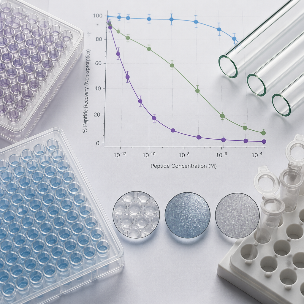
| Adsorption is concentration-dependent and most severe at low nanomolar concentrations | At sub-micromolar concentrations, the available surface area on labware walls can adsorb the majority of your peptide before the assay begins, producing a non-linear dose-response that mimics low potency. |
| Hydrophobic peptides lose signal linearity on standard polypropylene and borosilicate glass | Published data show only 10 to 20% peptide recovery from borosilicate glass and standard polypropylene tubes at 1 micromolar concentrations. Surface-modified low-bind vessels dramatically improve recovery. |
| BSA pre-coating at 0.5% (m/v) can block adsorption even at picomolar peptide concentrations | Overnight BSA coating followed by a water wash creates a competitive protein barrier on vial surfaces. This approach has been shown to prevent adsorption from solutions as dilute as 100 pM. |
| Low concentrations of surfactants (0.005 to 1%) reduce surface adsorption without harming assay biology | Tween-20, polyethylene glycol, and similar non-ionic surfactants interfere with hydrophobic interactions between peptide and surface. They must be kept out of mobile phases in LC-MS workflows to avoid ion suppression. |
| Peptide impurities introduce a second adsorption layer that high-purity compounds eliminate | Hydrophobic truncation fragments and deletion sequences from inadequate synthesis compete for binding sites and coat surfaces first, amplifying adsorption loss for the target peptide and contaminating binding assays. |
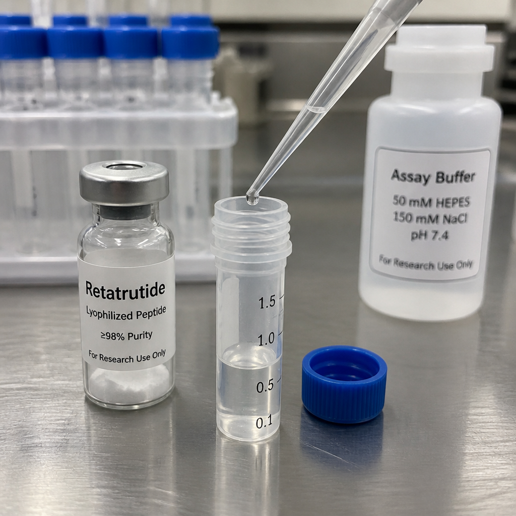
| Adding peptide directly to diluent, not to dry tube walls, reduces initial adsorption loss | Depositing concentrated peptide stock onto the side of an empty tube before adding buffer maximises surface contact time. Pipetting directly into buffer already in the vessel reduces that contact significantly. |
| Batch-specific COA and traceability allow you to isolate adsorption artefacts from compound variability | When you know the exact purity, counterion content, and moisture level of each vial from a verified batch, you can rule out compound-level variance as a cause of irreproducibility and focus on buffer and surface variables. |
What Is Peptide Non-Specific Adsorption and Why Does It Matter?
Non-specific adsorption (NSA), sometimes called non-specific binding (NSB), describes the loss of peptide analyte to surfaces it was never intended to contact: the walls of storage vials, pipette tips, autosampler components, microplate wells, and tubing. Unlike receptor-mediated binding, NSA is driven by physical chemistry, chiefly hydrophobic interactions, electrostatic attraction, and hydrogen bonding between the peptide and the surface material. The loss is silent, rapid, and often irreversible within the timescale of a typical assay setup.
The practical consequence is that the nominal concentration you calculated from your stock is not the concentration reaching your assay system. If your working concentration is 10 nM and 60% of that peptide has adsorbed to the plate and pipette tip before the incubation begins, you are dosing at an effective 4 nM. That shift moves you off the steep part of your concentration-response curve in ways that erode EC50 accuracy and reproducibility. Worse, the magnitude of adsorption is concentration-dependent: the fractional loss is largest at the lowest concentrations you test, which systematically bends the lower end of your curve and creates the illusion of a Hill slope anomaly or a weak binder.
Research examining peptide signal linearity across a dilution series has found that many peptides do not display a linear relationship between signal intensity and amount under standard conditions, and that increasing the organic content of the sample solvent improved linearity, pointing to concentration-dependent surface adsorption as the primary driver of the non-linearity. This is not an edge case: it is a routine feature of working with peptides in aqueous assay systems at low concentrations.
How Compound Purity Directly Drives Adsorption Risk
The relationship between compound purity and non-specific adsorption is not intuitive but it is direct. A peptide synthesised by solid-phase methods will typically carry deletion sequences, truncated fragments, incompletely deprotected intermediates, and in some cases enantiomeric impurities from racemisation during synthesis. These co-eluting or near-eluting species are not biologically inert bystanders.
Hydrophobic Impurities as Surface-Fouling Agents
Deletion sequences and truncation fragments, particularly those containing hydrophobic residues, preferentially adsorb to the same labware surfaces that adsorb your target peptide. They compete for the available surface sites and, in doing so, can either accelerate apparent loss of your active compound or, if they adsorb first and form a partial passivation layer, alter the adsorption kinetics in unpredictable ways. Hydrophobic impurities have been specifically identified as sources of non-specific signal in ELISA-type assays, where they compete for binding sites and alter the physical properties of the assay solution.
Enantiomeric Contaminants and Assay Interference
Racemisation during synthesis produces D-amino acid epimers that are mass-identical to the correct L-amino acid sequence. These enantiomers behave differently in biological systems and can produce anomalous binding signals. In a high-sensitivity binding assay, even a small percentage of D-epimer can shift an apparent Kd value or introduce a low-affinity population that broadens your competition curve. This class of impurity is invisible to purity calculations based on area under the curve alone unless the HPLC method specifically resolves enantiomers.
The practical implication is that a 99%+ purity specification verified by independent HPLC is not just a quality badge. It is a direct risk-reduction measure for both NSA-related artefacts and receptor-level interference. Every percent of impurity is a percent of material with unpredictable surface chemistry, unknown receptor affinity, and untested buffer interactions.
Pro tip: When you receive a new batch of lyophilised peptide, request a batch-specific COA that includes HPLC trace data, not just a purity percentage. A single-peak trace confirms you are not carrying forward a co-eluting impurity that the percentage alone would miss.
Assay Buffer Optimisation: The Variables That Actually Move the Needle
Buffer composition is the most accessible lever for controlling peptide non-specific adsorption in the assay environment. Most researchers inherit a buffer formulation and treat it as fixed, but for highly hydrophobic or amphipathic peptides, small compositional changes have measurable effects on both surface adsorption and assay signal quality.
Surfactants: Effective Concentrations and Compatibility Trade-offs
Non-ionic surfactants placed in the peptide-containing solutions, not in mobile phases, are among the most practical first-line mitigations. Tween-20, polyethylene glycol, and certain glucoside-based surfactants interfere with multiple surface interaction mechanisms simultaneously by inserting a steric barrier between peptide and surface. Published work on minimising peptide adsorption in assay contexts has shown that concentrations as low as 0.005% and up to approximately 1% are typically sufficient to meaningfully reduce adsorption for most peptide classes. Higher concentrations are not necessarily better and can begin to affect assay biology, particularly for cell-based receptor binding experiments. For LC-MS applications, surfactants must be kept out of the mobile phase entirely because they alter column selectivity and cause ion suppression.
Organic Modifiers and Their Role in Linearity
Adding an organic modifier, such as acetonitrile, to the peptide solution matrix has been shown to increase signal linearity across a dilution series by reducing surface adsorption. The mechanism is straightforward: reducing the polarity of the solvent environment diminishes the thermodynamic drive for hydrophobic peptides to escape to surfaces. The practical challenge in cell-based assays is that organic modifier concentrations above roughly 0.1% begin to affect cell viability, so their use is largely confined to analytical workflows like MS-based quantification rather than functional receptor assays.
pH and Ionic Strength Effects
Electrostatic contributions to adsorption depend on the charge state of the peptide and the surface. Adjusting buffer pH relative to the peptide isoelectric point changes the net charge and therefore the electrostatic component of adsorption. High ionic strength, for example through addition of sodium chloride at concentrations above 1.6 M, has also been investigated as a strategy to compete with electrostatic surface interactions. This approach is more useful in analytical preparation contexts than in functional assays, where high salt disrupts physiological receptor-ligand interactions.
Pro tip: When building a new assay buffer for a novel peptide, run a spike-and-recovery experiment at your lowest intended working concentration before investing in a full concentration-response series. Pipette your peptide into buffer in your actual assay vessel, incubate at assay temperature, then quantify what remains. If recovery at that concentration is below 80%, your buffer or vessel choice needs adjustment before any biological data will be interpretable.
Labware Surface Selection and Surface Passivation Strategies
Surface chemistry of labware is at least as important as buffer composition, and the choice of vessel type is often more impactful on peptide recovery than any buffer additive. This is especially true at working concentrations below 100 nM, where the surface-area-to-volume ratio of typical microplate wells and storage vials provides a proportionally large adsorption sink.
Container Material Performance
Standard borosilicate glass and standard polypropylene perform similarly poorly for hydrophobic peptide recovery. Research comparing container types has found that only 10 to 20% of peptide was recovered from borosilicate glass vials and standard polypropylene tubes at 1 micromolar concentrations after one hour of incubation, meaning 80 to 90% of the material was lost to the surfaces before any assay step began. Surface-modified low-bind containers, including certain specialty polypropylene formulations and polymer-coated plates, showed substantially better recovery for hydrophobic peptides in the same experiments.
Silanised glass vials represent a different strategy: deactivating the silanol groups on the glass surface reduces polar adsorption mechanisms. For hydrophobic peptides, however, silanised glass can still allow hydrophobic adsorption, so the choice of surface treatment must be matched to the dominant interaction chemistry of your specific peptide. A vial designed to reduce polar adsorption will not necessarily protect a hydrophobic peptide from hydrophobic surface loss.
BSA Pre-Coating as a Passivation Protocol
Pre-coating vials and assay vessels with bovine serum albumin (BSA) is one of the most reliable passivation strategies available and does not require specialised equipment. The protocol involves filling the vessel with 0.5% (m/v) BSA solution overnight at room temperature, then washing three times with water and drying before use. This BSA layer physically occupies the high-energy adsorption sites on the surface. Research has confirmed that this pre-coating step can prevent adsorption of peptides from solutions as dilute as 100 pM. A key distinction applies: pre-coating the surface prior to adding the peptide is more effective than simply including BSA in solution alongside the peptide, though the latter approach also provides measurable benefit by reducing the effective free surface area available for adsorption.
Pipetting Technique as an Adsorption Variable
Technique-level decisions matter more than most researchers realise. Pipetting peptide stock directly onto dry tube walls before adding buffer maximises the contact time between concentrated peptide and bare surface. Pipetting the stock directly into buffer already present in the vessel substantially reduces that initial high-surface-contact event. Pre-rinsing pipette tips several times with the peptide solution before aspirating the final volume is a simple, cost-free step that significantly reduces adsorptive loss to the tip interior. Using a fresh tip for each serial dilution prevents carryover that can mimic concentration-dependent effects in your data.
Comparison of Non-Specific Adsorption Mitigation Approaches
| Mitigation Approach | Best Use Context | Key Limitations |
|---|---|---|
| BSA surface pre-coating (0.5% m/v overnight) | ELISA, functional binding assays, storage vials for sub-nanomolar working stocks. Effective even at 100 pM peptide concentrations. | Adds a preparation step before each experiment. BSA must be compatible with downstream detection (can interfere with certain antibody-based readouts). Not suitable for LC-MS mobile phases. |
| Surfactant addition to peptide buffer (0.005 to 1% Tween-20 or PEG) | Aqueous assay buffers, peptide dilution series, microplate-based receptor binding assays. Practical first-line intervention. | Must be excluded from LC-MS mobile phases. At higher concentrations can affect cell viability and receptor biology. Does not address all interaction types equally. |
| Surface-modified low-bind labware (specialty polymer coatings) | All peptide work below 1 micromolar, particularly for hydrophobic sequences. Provides structural solution rather than chemical masking. | Higher cost per vessel. Not all "low-bind" products perform equivalently across peptide classes. Some commercial low-retention tubes can paradoxically increase adsorption for certain proteins and peptides, so empirical verification for your specific compound is required. |
Retatrutide-Specific Considerations in Receptor Binding Assays
Retatrutide (LY3437943) is a triple agonist targeting GLP-1, GIP, and glucagon receptors. Its pharmacological profile makes it a valuable compound for in vitro research into metabolic regulation, insulin sensitivity, and energy balance signalling. From an adsorption standpoint, its structural features, including the lipidic acyl chain modification present in fatty acid-conjugated GLP-class peptides, place it firmly in the category of peptides at high risk for hydrophobic surface adsorption.
The fatty acid side chain that contributes to its pharmacokinetic properties in physiological contexts also gives it a strong affinity for hydrophobic surfaces in an aqueous assay environment. This means that a standard polypropylene microplate or a glass storage vial without surface treatment will adsorb a significant fraction of your high purity retatrutide working stock, particularly at the nanomolar concentrations relevant to receptor binding assays. The nominal concentration on your dilution scheme will not reflect the actual concentration in your wells.
For receptor binding studies with retatrutide, the combination of low-bind assay plates, buffer containing a low concentration of non-ionic surfactant compatible with your detection method, and a pre-rinsed pipette tip protocol is the recommended starting configuration. Because retatrutide's triple receptor activity means you may be working across multiple cell lines or membrane preparations with different basal binding characteristics, controlling NSA is especially important: variability from adsorption will compound with the inherent variability of multi-receptor assay systems and make cross-system comparisons unreliable.
In peptide research, the difference between a reproducible assay and an irreproducible one is often found not in the biology but in the gap between the nominal concentration you intended to apply and the actual concentration that reached your system. Closing that gap is a pre-analytical problem, not a statistical one.
Starting with a verified 99%+ purity lyophilised compound from a supplier that provides independent Canadian laboratory verification and full chain of custody documentation is the first step in closing that gap. Compound variability and impurity-driven adsorption are not controllable after the vial is opened. Assay buffer composition and labware selection are. But they can only do their job if the input material is clean enough that you are only solving one adsorption problem at a time.
Chain of Custody and Batch Traceability as Data Integrity Tools
In the context of protein binding and peptide non-specific adsorption research, batch traceability is a practical tool, not merely a compliance formality. When assay results shift between experiments, there are three candidate explanations: biological variation in the assay system, buffer or labware changes, and variation in the compound itself. Without verified, batch-specific documentation on your peptide, you cannot isolate which variable is responsible.
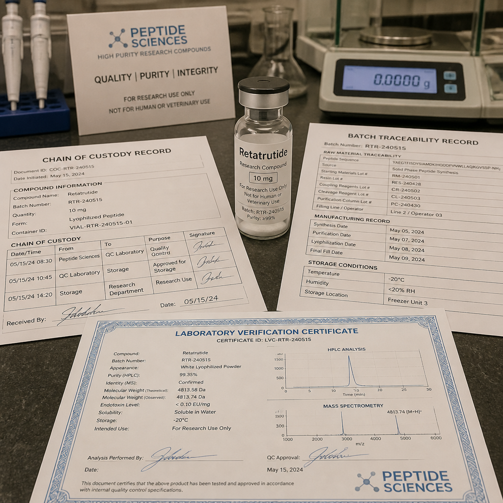
A chain of custody document paired with a batch-specific COA from independent laboratory analysis gives you a fixed reference point for compound identity, purity, and counterion content. If purity is consistent across batches and your assay results still shift, you can direct your troubleshooting toward the buffer and surface variables discussed in this article. If purity shifts, you have identified the source of variance before wasting time on buffer reformulation that will not solve a compound-level problem.
For research institutions managing multiple investigators, peptide lot-to-lot traceability also provides a practical audit trail when reviewing data. Knowing that two experiments used different lot numbers of the same peptide, with documented purity differences, transforms an apparent inconsistency in the data into a traceable and explainable finding. The alternative, working from undocumented material, produces discrepancies with no clear resolution path.
Pepura Labs provides lyophilised retatrutide and other research-grade compounds with full chain of custody documentation and independent Canadian laboratory verification. Researchers sourcing for in vitro applications can review available compounds and documentation at pepuralabs.ca/shop.
Frequently Asked Questions
What is the single most effective step to reduce peptide non-specific adsorption in a cell-based receptor assay?
Switching to a surface-modified low-bind assay plate and pre-rinsing pipette tips with your peptide dilution before aspirating your final working volume will together reduce NSA more than any buffer additive alone. If you can only change one variable, switch your vessel type first, because the surface area of a standard microplate well represents a substantial adsorption sink relative to the working volume of a typical assay well.
Does using a carrier protein like BSA in the assay buffer interfere with receptor binding?
It depends on the assay format and the BSA concentration. In functional GPCR assays with intact cells, low concentrations of BSA (0.1 to 0.5%) are commonly used and generally do not abolish receptor activity, but they do reduce the free fraction of highly albumin-binding peptides. For peptides with strong serum albumin affinity, such as fatty-acid-conjugated sequences, including BSA in the assay buffer can shift your apparent EC50 to higher concentrations by reducing the freely available peptide. This is distinct from true potency loss and must be accounted for in data interpretation.
Why does my dose-response curve look normal at high concentrations but miss the bottom plateau?
This is a classic signature of concentration-dependent surface adsorption. At high concentrations, the available surface sites become saturated with peptide, and enough free peptide remains in solution to produce a near-maximal response. At low concentrations, the surface-to-analyte ratio means a larger fraction adsorbs, which removes the signal from the bottom of the curve. The result is an apparently truncated curve with a displaced EC50. Improving surface passivation and buffer conditions specifically benefits the low-concentration end of your dilution series and should restore the missing plateau.
Is it safe to include Tween-20 in a radioligand binding assay buffer?
Low concentrations of Tween-20 (typically below 0.05%) are routinely used in radioligand binding buffers and are generally compatible with the assay format. At higher concentrations, non-ionic detergents can solubilise membrane components and disrupt receptor presentation, which would invalidate the assay. For any new buffer formulation, run a control experiment comparing Bmax and Kd values with and without the surfactant at your intended concentration before committing to its inclusion as a standard formulation step.
How does peptide purity affect non-specific adsorption specifically, as opposed to just receptor binding specificity?
Impurities, particularly hydrophobic deletion sequences and truncated fragments from solid-phase synthesis, occupy surface adsorption sites and alter the apparent adsorption kinetics of the target peptide. They can pre-coat surfaces in a way that either accelerates or partially blocks subsequent target peptide adsorption, and this effect is concentration-dependent and batch-variable. Starting with a 99%+ purity compound verified by independent HPLC removes this layer of unpredictability, so your buffer and surface optimisation work addresses the single target compound rather than a mixture with compound-specific surface chemistry you cannot fully characterise.
Can aggregation of a lyophilised peptide contribute to false non-specific adsorption signals?
Yes. Improper reconstitution of lyophilised peptide can produce aggregates that behave differently from monomeric peptide: they may adsorb to surfaces more rapidly, pass through pipette tips inconsistently, or produce anomalous activity in functional assays. Reconstituting in a compatible aqueous solvent, using gentle mixing rather than vigorous vortexing for aggregation-prone sequences, and performing a brief low-speed centrifugation to remove insoluble aggregates before preparing your working dilution series are standard steps that reduce aggregate-related artefacts. Batch-specific documentation showing moisture content and the lyophilisation conditions provides useful context for reconstitution choices.
How do I verify that my assay buffer changes have actually reduced non-specific adsorption rather than just changing my assay signal?
Run a spike-and-recovery experiment independent of your biological assay. Prepare a peptide solution at your lowest intended assay concentration in your test buffer and vessel, incubate at assay temperature for the duration of your assay, then quantify the remaining peptide by HPLC, LC-MS, or a validated immunoassay depending on your available instrumentation. Compare recovery to a freshly prepared sample that was not incubated in the vessel. A recovery above 80% at your minimum working concentration is a reasonable threshold before investing in full assay characterisation. This experiment should be repeated whenever you change buffer formulation, vessel type, or peptide batch.
Have you run into non-specific adsorption problems that sent you down an unexpected troubleshooting path? Share what you found in the comments or reach out via social, we would like to hear how different research groups are approaching this problem in practice.
References
- Recommendations for peptide generation, quantification, storage and handling for mass spectrometry-based assays (PMC4830481)
- Tutorial review on pre-analytical considerations for peptide assays: buffer and handling optimisation strategies
- Study on cationic peptide adsorption to glass and polypropylene surfaces and the effect of container choice on peptide recovery
- Research on the effect of peptide surface adsorption on signal linearity and approaches to improve quantification reliability
- White paper on achieving maximum protein and peptide recovery by minimising non-specific binding in analytical workflows