Hepatic fat accumulation is one of the least forgiving endpoints in metabolic research. If your in vitro model does not accumulate lipid in a way that resembles real liver tissue, every downstream reading from a retatrutide glucagon study becomes hard to interpret. Non-alcoholic fatty liver disease affects roughly 1 in 4 adults according to a widely cited review, which is why laboratories keep building cell-based systems to study it. The real question is not whether to use a hepatic fat in vitro model, but which liver cell line will give you a defensible answer. This guide explains how we approach that choice at Pepura Labs, where we supply research-grade retatrutide compounds to laboratories across Canada.
Table of Contents
- Quick Takeaways
- Why Hepatic Fat Models Matter for Retatrutide Research
- What a Useful In Vitro Hepatic Fat Model Must Do
- Comparing Liver Cell Lines for Hepatic Fat Studies
- Glucagon Receptor Biology in Hepatocyte Models
- Lipid Loading Protocols and Vehicle Controls
- Readouts and Verification for Steatosis Assays
- Common Selection Mistakes and How to Avoid Them
- Frequently Asked Questions
- References
Quick Takeaways
| Key Insight | Explanation |
|---|---|
| HepG2 is the common baseline, not automatically the right answer | Most published 2D NAFLD work uses HepG2, which helps comparability with earlier studies but limits some metabolic questions. |
| Primary hepatocytes remain the reference standard | They hold metabolic function best, but donor variability and short culture windows make multi-week lipid studies harder to run. |
| Match the model to the glucagon question | Amino acid catabolism and glucose output readouts require a line with confirmed glucagon responsiveness, not an assumed one. |
| Confirm lipid loading before testing the compound | Fatty acid loading should produce measurable neutral lipid accumulation in vehicle controls before any retatrutide arm is interpreted. |
| Pair steatosis readouts with viability and stress markers | Lipid staining alone can be confounded by toxicity, so run viability and oxidative stress panels on the same plates. |
| Log batch, COA, and reconstitution details for every run | Compound identity, purity, and reconstitution solvent such as bacteriostatic water should be recorded per experiment for reproducibility. |
Why Hepatic Fat Models Matter for Retatrutide Research
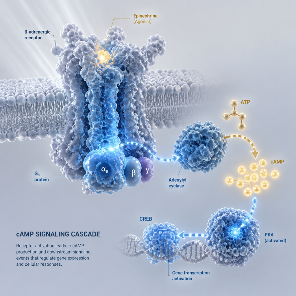
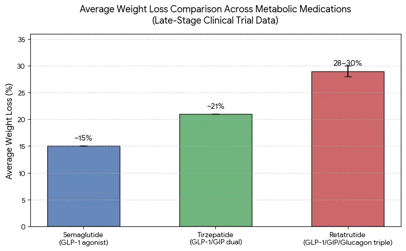
Retatrutide is a triple agonist that acts on GIP, GLP-1, and glucagon receptors. In a liver context, the glucagon arm changes the conversation, because glucagon signaling influences hepatic amino acid catabolism and glucose output, and those pathways feed back into how the liver handles lipid. A glucagon study that ignores fat handling is only telling part of the story.

Fatty liver is the second most widespread liver disease after viral hepatitis, and its incidence is rising. That makes the hepatic fat in vitro model more than a niche tool. It is the bridge between a receptor-level observation and a tissue-level claim that a reviewer will eventually ask you to defend.
What Fat Accumulation Tells You That Glucose Output Does Not
Glucose output assays are fast and sensitive, but they capture a snapshot of one pathway. Steatosis readouts, such as neutral lipid staining and triglyceride quantification, show whether the cells are storing energy in a way that resembles a fatty liver. When a compound shifts glucose output but leaves lipid accumulation untouched, that is a finding worth reporting, not a gap to hide.
Pro tip: Run glucose output and lipid accumulation on the same seeding density and time course. Comparing numbers from separate plates is one of the fastest ways to create an artifact that looks like a biological effect.
Why Retatrutide Makes the Model Choice Harder
Because retatrutide engages several incretin and glucagon-related receptors at once, a simple readout can reflect a mix of direct and indirect effects. A well-chosen cell line helps you separate those contributions. A poorly chosen one can make every result look plausible while none of them are clearly attributable.
At Pepura Labs, we supply lyophilized retatrutide in research strengths of 10mg, 20mg, 40mg, 60mg, and 84mg, each with a certificate of analysis and batch traceability. Those documents only help if your own records link each batch to each plate, so treat them as part of the experimental design rather than paperwork you file later.
What a Useful In Vitro Hepatic Fat Model Must Do
Before comparing specific cell lines, it helps to define what a usable model must accomplish. A hepatic fat in vitro model should accumulate lipid under a defined stimulus, respond to the signaling pathway you care about, and remain stable enough across passages to support a meaningful comparison. If a model fails any one of those tests, the rest of your data is harder to trust.
The review literature groups the main model types into primary hepatocytes, immortal cell lines, hepatocyte-like cells derived from stem cells, and precision-cut liver slices. Each category trades off physiological fidelity, throughput, and cost. Culture format matters too, since monoculture, co-culture, and three-dimensional systems change how lipid and signaling behave.
Lipid Accumulation That Reflects Steatosis
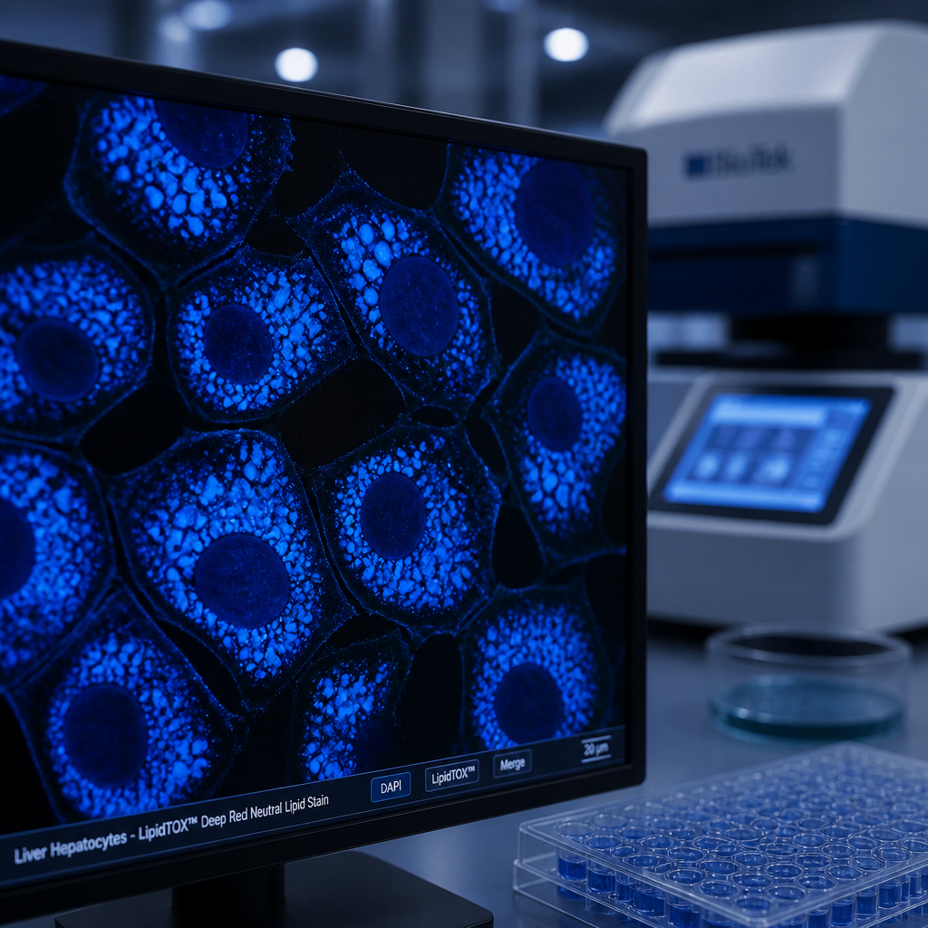
A useful model stores lipid in droplets that are visible, quantifiable, and responsive to fatty acid loading. If a line shows almost no neutral lipid even after loading, it may still be useful for signaling studies, but it is a poor choice for a steatosis endpoint. Set that expectation before you start, not after the first disappointing plate.
Responsiveness to Glucagon Signaling
Glucagon responsiveness cannot be assumed from the cell name. You need a functional readout, such as a cAMP response or a downstream metabolic change, that confirms the pathway is active in your specific culture. Without that check, a null result may reflect a silent receptor rather than a true absence of effect.
Stability Across Passages and Batches
Immortal lines are convenient because they are reproducible in principle, but drift over passages can change morphology, lipid handling, and receptor expression. Record passage number for every experiment, and avoid comparing data generated at very different passage ranges as if they were the same model.
Comparing Liver Cell Lines for Hepatic Fat Studies
The three options most labs weigh for a hepatic fat in vitro model are HepG2 cells, HepaRG cells, and primary human hepatocytes. Each has a legitimate use case, and each has a limitation that will show up in your data if you ignore it. The right choice depends on whether your question is about comparability, metabolic depth, or physiological realism.
HepG2 Cells: The Common Baseline
HepG2 is the most common immortalized cell line used for two-dimensional in vitro models of NAFLD. That widespread use is its main advantage, because it lets you compare your results with a large body of published work. It is inexpensive and easy to maintain, which suits screening-scale experiments.
The weakness is that immortal hepatoma-derived cells do not behave like mature hepatocytes in every respect. Drug-metabolizing capacity and some metabolic pathways are often weaker, so interpret amino acid and metabolic readouts with that limitation in mind.
HepaRG Cells: A Closer Metabolic Profile
HepaRG cells are often chosen when labs want a metabolic profile closer to primary tissue than HepG2 offers. Comparative studies have examined both lines side by side for modeling liver metabolism, and the differences between them are large enough to change conclusions. If your glucagon study depends on amino acid handling, HepaRG deserves a direct head-to-head test before you commit.
The trade-off is cost, culture time, and a smaller body of directly comparable literature. Plan for longer differentiation timelines and budget accordingly.
Primary Hepatocytes: The Reference That Is Hard to Scale
Primary hepatocytes are widely described as the gold standard for studying hepatic glucose, lipid, and cholesterol metabolism in vitro. When a reviewer asks whether a finding holds in a more physiological system, primary cells are the answer they expect. The cost is donor-to-donor variability, limited culture life, and difficulty running large experiments.
The table below summarizes how these options compare for hepatic fat and glucagon work.
| Cell model | Strengths for hepatic fat work | Limitations for glucagon studies |
|---|---|---|
| HepG2 cells | Widely published, inexpensive, easy to expand, good for comparability with earlier steatosis work | Weaker metabolic and drug-handling profile, so amino acid and glucose output effects need careful interpretation |
| HepaRG cells | Closer metabolic profile, useful for head-to-head comparison with HepG2 in metabolism studies | Longer culture and differentiation timelines, smaller direct literature base, higher cost per experiment |
| Primary hepatocytes | Reference standard for hepatic glucose, lipid, and cholesterol metabolism, strongest physiological relevance | Donor variability, limited culture window, difficult to scale across large or repeated experiments |
Glucagon Receptor Biology in Hepatocyte Models
Glucagon acutely regulates hepatic amino acid catabolism, and this has been tested directly in immortal hepatocyte lines such as HepG2 and HepaRG. That work shows the pathway can be measured in these systems, but it also shows that the response is not identical across lines. Treat each cell line as its own biological system rather than a stand-in for the liver.

A practical rule is to validate the glucagon response before running any retatrutide experiment. Use a known glucagon reference treatment on the same plates, and confirm the readout moves in the expected direction. If the reference does not respond, you have a model problem, not a compound result.
Confirming Receptor Expression Before You Test
Receptor expression should be checked at the start of a project and again after major passage changes. Transcript-level or protein-level confirmation is more informative than assuming expression from the literature. Keep the confirmation data in the same record as your experiment so reviewers can see what was verified.
Separating Glucagon Effects From Lipid Effects
Glucagon signaling and fatty acid loading both change hepatocyte metabolism, so their effects can overlap. Build a design where glucagon alone, lipid alone, and the combination each have their own arms. Without that structure, you cannot say whether a change in lipid handling came from the receptor pathway or from the loading protocol.
A model that cannot show its own glucagon response is a model you have not yet validated, no matter how many plates you have run through it.
Lipid Loading Protocols and Vehicle Controls
Most steatosis models rely on loading cells with fatty acids, typically oleate, palmitate, or a mixture, to drive intracellular lipid accumulation. The loading step is where many experiments quietly fail. A protocol that works for one line may overload another, causing toxicity that looks like a compound effect.
Start with a loading titration on your chosen cell line. Measure lipid accumulation and viability at several fatty acid concentrations and time points, then pick a condition that produces clear steatosis without large losses in cell number. That pilot run is not optional if you want the main experiment to be interpretable.
Choosing a Fatty Acid Load
Oleate tends to produce neutral lipid storage with less toxicity than some saturated fatty acids, which makes it a common starting point for steatosis work. Palmitate can induce stress responses that complicate interpretation. Whatever you choose, state the species, concentration, and vehicle in the methods so another lab can reproduce the model.
The MDPI study on HepG2 cells used lipid accumulation and oxidative stress together as a platform for evaluating therapeutic effects, which is a sensible design pattern to copy. Measuring stress alongside storage tells you whether a lipid change is protective, neutral, or damaging.
Building Vehicle and Time Controls
Every plate needs a vehicle control that matches the solvent and concentration used for the compound. Retatrutide is reconstituted and diluted before use, so vehicle composition, including any bacteriostatic water and dilution steps, must be identical across arms. Include time-matched controls as well, because lipid accumulation changes with culture duration even without treatment.
Pro tip: Freeze a single reconstituted aliquot per batch for the whole experiment whenever stability allows, and record the thaw count. Variation between aliquots is an easy source of batch-to-batch noise that looks like biology.
Readouts and Verification for Steatosis Assays
A steatosis claim is only as strong as the readouts behind it. Neutral lipid staining is quick and visual, but it is semi-quantitative and sensitive to imaging conditions. Triglyceride extraction and quantification give a bulk number, though they lose spatial information. Using both is the most defensible approach when the result will be published or shared with a partner.
Pair every lipid readout with a viability assay and, where possible, an oxidative stress marker. A compound that reduces lipid by killing cells is not a clean finding. Report the viability numbers beside the lipid numbers, even when they look unremarkable.
Choosing Between Staining and Extraction
Staining works well for screening and for showing where lipid sits within the cell. Extraction works better when you need a number that can be compared across plates and runs. A common mistake is using staining for a primary endpoint and then trying to compare intensities between experiments imaged on different days.
Documenting Compound Identity for Each Run
Each run should record the compound batch number, the certificate of analysis reference, the reconstitution date, and the diluent used. Our batches are independently verified by Canadian laboratories with full chain of custody documentation, but that verification only helps your study if the batch number travels with your data. Store the paperwork alongside the raw plate files.
Common Selection Mistakes and How to Avoid Them
Most poor model choices come from convenience rather than from a deliberate match between the question and the system. A line gets chosen because it is already in the freezer, or because a previous project used it, and nobody checks whether it fits the glucagon readout. The result is a dataset that is internally consistent but hard to defend.
The fix is to write down the question first. Specify whether you need comparability, metabolic depth, or physiological realism, then choose the model that best serves that priority. If the answer changes midway through the project, that is a signal to re-validate, not to quietly switch lines.

Choosing a Cell Line for Convenience Alone
Convenience is a valid factor, but it should not be the only one. A line that is easy to grow may still have a weak glucagon response or a poor lipid phenotype. Run a short pilot comparing two candidate lines on your core readouts before committing a full study budget.
Skipping Passage and Donor Documentation
Passage number, cell source, and donor details are often missing from methods sections, and that omission makes results hard to reproduce. Keep a simple log for each culture, including thaw date and passage count. When you compare runs months apart, that log is what tells you whether a difference is biological or procedural.
Frequently Asked Questions
Which cell line should I start with for a hepatic fat in vitro model?
For most first-pass studies, HepG2 is a practical starting point because of its wide use in published NAFLD work. Run a small pilot against HepaRG or primary cells if your glucagon readouts depend on amino acid or metabolic handling.
Are primary hepatocytes always better than immortal cell lines?
They are closer to physiological liver tissue, but they are not always better for your question. Donor variability and short culture windows can make them a poor choice for large, repeated, or long-duration experiments.
How do I confirm that my cells respond to glucagon?
Include a known glucagon reference treatment on the same plates and check that a functional readout moves in the expected direction. Confirm receptor expression at the start of the project and after major passage changes.
What lipid loading controls are essential for a steatosis assay?
Use vehicle controls matched to every compound arm, time-matched controls, and a viability readout on the same plates. Without these, you cannot separate lipid storage from toxicity or solvent effects.
Why does batch documentation matter in cell-based retatrutide research?
Batch numbers, certificates of analysis, and reconstitution details let you trace each result back to a specific compound lot. That traceability is what makes a finding reproducible when a colleague or reviewer asks how the material was prepared.
Which liver cell line has given your lab the most reliable steatosis data, and what would you change in your current protocol?
References
- Review of in vitro models for non-alcoholic fatty liver disease and their emerging platforms
- Overview of molecular developments in cell models of fatty liver disease
- Study of acute glucagon effects on amino acid catabolism in HepG2 and HepaRG cells
- Comparison of HepaRG and HepG2 cell lines for modeling liver metabolism
- Study of lipid and glucose metabolism in hepatocyte cell lines compared with primary hepatocytes