If your pancreatic beta cell assay gives a clean insulin curve on Monday and a flat response on Thursday, the compound is rarely the first suspect. The assay is. Glucose-stimulated insulin secretion looks simple on paper: hold cells at low glucose, switch to high glucose, measure insulin. In practice, small differences in cell density, buffer make-up, and pre-incubation timing can bury the signal you came to measure. This guide walks through how we approach beta cell assay design for GLP-1 receptor research, where the GLP-1 mechanism enters the picture, and which controls separate a real secretory effect from noise.
Table of Contents
- Quick Takeaways
- Why Beta-Cell Biology Sets the Assay Rules
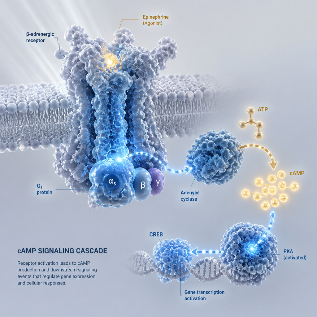
- GLP-1 Receptor Signaling in Beta Cells
- Designing the Glucose-Stimulated Insulin Secretion Assay
- Comparing Insulin Secretion Assay Formats
- Controls, Viability, and Apoptosis Readouts
- Common Mistakes in Insulin Secretion Data
- Sourcing and Documenting Research Compounds
- Frequently Asked Questions
- References
Quick Takeaways
| Key Insight | Explanation |
|---|---|
| Insulin output is the primary endpoint | Insulin is the only hormone that lowers blood glucose, so secreted insulin should anchor every beta cell assay, with viability as a supporting measure. |
| Separate short-term secretion from long-term survival | A few hours of exposure tests secretory function. Apoptosis and survival effects need longer exposure windows and their own readouts. |
| Map alpha-cell contributions before adding a GLP-1 agonist | Pancreatic alpha cells can produce active GLP-1, so islet preparations may generate their own incretin signal before any compound is added. |
| Run a receptor antagonist arm | An antagonist arm tells you whether an effect runs through the GLP-1 receptor or through another pathway. |
| Normalize insulin to cell content | Normalizing to DNA or protein content reduces noise from uneven cell loading across wells. |
| Document the compound lot before you run the plate | Lot-level certificates of analysis and batch traceability let you tie a shifted result to a specific material. |
Why Beta-Cell Biology Sets the Assay Rules
The pancreatic beta cell has one job that matters most for blood sugar: it secretes insulin in proportion to glucose. A 2017 review by P. Rorsman describes the beta cell as central to glucose homeostasis and notes that insulin is the only hormone capable of lowering blood glucose concentration. That single-hormone role is why insulin release, not cell count or morphology alone, should anchor most beta cell assays.

Glucose sensing and electrical activity
The title of that 2017 review points to electrical activity as the link between glucose and secretion. Glucose metabolism changes the beta cell's membrane electrical activity, which drives calcium entry and the release of insulin-containing granules. If your protocol measures insulin only at the end of a long window, you see the integrated result of that chain, not its kinetics.
Insulin release as a metabolic readout
A 2012 metabolomic study by M. Huang examined beta-cell insulin release as a response to rising nutrient levels. The practical lesson is that insulin output reflects upstream metabolism, so a flat secretion curve can come from a metabolic problem in the cells rather than a failed receptor. When a result is unexpected, pair insulin with a metabolic readout before you blame the compound.
Pro tip: Report basal and stimulated insulin as separate values. A fold-change ratio can look healthy while basal release has quietly climbed, and that changes how you read any GLP-1 effect.
GLP-1 Receptor Signaling in Beta Cells
GLP-1 receptor agonists act on beta cells through the GLP-1 receptor, and the classic effect is amplification of glucose-dependent insulin release. A 2025 systematic review by N. Rea included seven preclinical studies on GLP-1 receptor agonists and beta-cell apoptosis. It reported a robust anti-apoptotic effect in those models, which is a separate question from secretion.
Secretion and survival are different endpoints
A secretion assay that runs for a few hours tests how cells respond right now. Survival effects take longer to show and need their own design, such as extended culture under a defined stressor. Mixing these up is the fastest way to write a conclusion the data cannot support.
Where retatrutide fits in the pathway
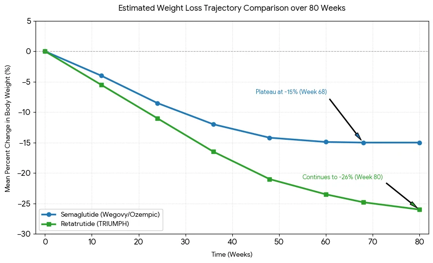
Retatrutide is a multi-receptor agonist, so its GLP-1 component cannot be assumed to be the only signal in your well. It engages the GLP-1 receptor alongside the GIP and glucagon receptors, which means a response may reflect more than one receptor. A 2021 review by S. E. Alavi covers GLP-1 receptor agonists such as exendin-4 and sequence modifications designed to target beta cells, a useful frame for deciding what your compound does at the receptor level.
Coverage of recent work on pancreatic alpha cells reports that they can produce active GLP-1. That means islets are not a blank slate, and some of the incretin background in your preparation may be self-generated before any compound is added.
Pro tip: Run a GLP-1 receptor antagonist arm alongside the agonist arm. Exendin (9-39) is a commonly used antagonist. If the effect survives antagonism, you are looking at a pathway outside the GLP-1 receptor, and your mechanistic conclusion changes entirely.
Designing the Glucose-Stimulated Insulin Secretion Assay
A glucose-stimulated insulin secretion assay has three moving parts: a basal condition, a stimulatory condition, and a readout window. Each needs to be set deliberately for your cell model rather than copied from a protocol written for a different system. We validate glucose concentrations and incubation times in pilot runs before any compound enters the design.

Cell model and pre-incubation
Pre-incubation in low-glucose buffer clears residual glucose and lets secretion settle to baseline. Skip it, and the basal value carries the previous step's glucose, which compresses your apparent stimulation. Keep the buffer composition, including bicarbonate and HEPES, consistent across runs so that a buffer change does not look like a biological change.
Islets, dispersed islet cells, and cell lines respond differently to the same protocol. Treat each as a separate validation problem, and do not transfer a stimulation window from one system to another without re-running the glucose response first.
Timing, replicates, and normalization
Use enough replicate wells to detect a shift of the size you care about, and run every treatment at matched times across all plates. Normalize insulin to DNA or protein content so that well-to-well differences in cell loading do not masquerade as a pharmacological effect. Replicate across days, not only across wells.
In a mixed islet preparation, part of the incretin signal may be self-generated, so the baseline has to be measured before the agonist goes in.
Comparing Insulin Secretion Assay Formats
No single format answers every question. Static batch assays are simple and easy to scale, while perifusion systems show secretion kinetics that a single endpoint hides. Cell-line and stem-cell-derived models offer consistency but may not reproduce the full islet response. The table below sets out the trade-offs we weigh before choosing a format.
| Assay format | Best use in GLP-1 work | Main limitation |
|---|---|---|
| Static batch GSIS on isolated islets | Endpoint comparison of secretion across glucose levels and antagonist arms | A single time point hides kinetics, and islet preparations vary by donor and isolation |
| Perifusion system with isolated islets | Measuring first-phase and second-phase kinetics after a GLP-1 agonist challenge | Higher equipment demand and lower throughput |
| Beta-cell line or stem-cell-derived beta-like cells | Higher-throughput screening and run-to-run reproducibility | May not reproduce the full islet response, so key findings need confirmation in islets |
In practice, we use the static format for screening and move hits to perifusion when the kinetic question matters. Confirming a finding in a second system is what separates a result from a pattern. A result that appears in only one format should be described as preliminary in any write-up.
Donor variability is the hidden cost of primary islets. Two preparations from different donors can differ in their cellular composition, which feeds directly into the GLP-1 background discussed above. Record the isolation lot and islet count for every run so you can see that variability rather than average it away.
Controls, Viability, and Apoptosis Readouts
Every plate needs a vehicle control, a positive secretagogue control, and a no-glucose baseline at minimum. Without the positive control, a flat compound response cannot be distinguished from a dead assay. A Diabetes journal evaluation compared widely used beta-cell function indices measured under basal conditions and after glucose ingestion, which supports reporting basal and stimulated values separately.

Viability as a separate measurement
Measure viability in the same wells or matched wells, using a method that does not interfere with insulin measurement. A cell that secretes less may simply be dying, and the insulin number alone cannot tell you which. Pair the secretion data with a viability readout in every run.
Apoptosis needs its own design
Apoptosis is a longer story. The 2025 review by Rea reported anti-apoptotic effects of GLP-1 receptor agonists in preclinical models, but the studies it covered used different stress models and endpoints. When you test survival, define the stressor, the exposure window, and the apoptosis marker before you collect data, not after.
Antagonist and vehicle arms
The antagonist arm should run with the same agonist concentration and timing as the active arm. Compare the antagonist-only group with vehicle as well, because an antagonist can alter baseline secretion on its own. Without that comparison, a shift can be misread as a receptor effect.
Common Mistakes in Insulin Secretion Data
Most failed beta cell assays fail for mundane reasons. The most common is inconsistent timing, so the first and last wells in a plate differ by more than the treatment effect. Standardize pipetting order and timing from the start, and write them into the protocol.
Reading a single time point as a curve
A single insulin number treats secretion as a snapshot. A GLP-1 effect may show up early and fade, or appear only after a defined pre-incubation. If budget limits you to one time point, say so explicitly in your write-up and avoid kinetic claims.
Confusing the diluent with the compound
Solvents and diluents carry their own biology. Check that your vehicle matches the final concentration in every treated well, and check whether the preservative in your diluent is compatible with your cell system. A bacteriostatic diluent that works for reconstitution may still be the wrong choice for a sensitive culture.
Overreading small differences is the last trap. A difference visible inside one plate is a hypothesis, not a finding, until it repeats on a different day with a different plate.
Pro tip: Keep a plate map with lot numbers, passage numbers, and reagent lot IDs in the same file as the raw data. When a curve shifts between runs, the map is usually the fastest route to the cause.
Sourcing and Documenting Research Compounds
Assay results are only as reliable as the material behind them. For research use only, we supply retatrutide as a lyophilized peptide in 10 mg, 20 mg, 40 mg, 60 mg, and 84 mg compound formats. Our products are specified at 99% purity or higher and are independently verified by Canadian laboratories, with Chain of Custody documentation.
What to check on a certificate of analysis
Look for the lot number, the analytical method, the purity result, and the date of testing. Confirm that the lot on the certificate matches the vial in your hands, and link that record to your assay file. Batch traceability is what lets you retest a suspect lot instead of discarding an entire series.
Handling lyophilized material before the assay
Store lyophilized material according to the supplier's instructions and reconstitute it under controlled conditions. Prepare aliquots so that one vial is not repeatedly exposed to temperature swings and air. Record the reconstitution date and diluent lot alongside the assay data.
Canadian laboratory testing and documented chain of custody do not replace your own controls, but they remove one large source of uncertainty. You can then spend your effort on the biology. You can browse our current range at Pepura Labs.
Frequently Asked Questions
What is a pancreatic beta cell assay used for?
It measures how beta cells respond to a stimulus, most often by quantifying insulin secretion under low and high glucose. Researchers use it to study secretagogues, incretin agonists, and related compounds in vitro.
How does GLP-1 increase insulin secretion in vitro?
GLP-1 receptor activation potentiates glucose-dependent insulin release, so the effect is generally strongest when glucose is high and weak when it is low. Confirm this in your own model with a glucose-response curve and an antagonist arm.
Why does a retatrutide study need a receptor antagonist control?
Retatrutide acts on more than one receptor, so a response may not come from the GLP-1 receptor alone. An antagonist arm tests whether the GLP-1 receptor accounts for the effect you observe.
Is glucose-stimulated insulin secretion the same as beta-cell function?
No. GSIS is one readout of secretory capacity. Beta-cell function also includes survival, gene expression, and metabolic coupling, so pair GSIS with at least one additional measure before drawing functional conclusions.
What documentation should I keep for a research compound?
Keep the lot number, certificate of analysis, purity result, verification details, and chain of custody record, and link them to each assay run. This lets you trace a result back to a specific material if the data shift later.
What is the most stubborn step in your insulin secretion protocol, and how did you resolve it?
References
- PubMed summary of a systematic review on GLP-1 receptor agonists and beta-cell apoptosis
- Full-text article reviewing preclinical studies of GLP-1 receptor agonists on beta-cell apoptosis
- Review of pancreatic beta-cell electrical activity and insulin secretion
- Critical evaluation of indices used to assess beta-cell function in Diabetes
- Summary of research showing pancreatic alpha cells produce active GLP-1