Retatrutide serum protein binding is one of the most overlooked variables in cell culture work. Most protocols quietly assume that the concentration written on the tube is the concentration the cells actually see. That assumption breaks as soon as serum enters the medium. Retatrutide is a triple agonist that acts on GLP-1, GIP, and glucagon receptors, and like many peptides designed for longer exposure, it can interact with plasma proteins. Only the unbound portion is generally available to engage receptors in a simple equilibrium model, so this guide explains how to estimate the free fraction and calculate an honest effective concentration for in vitro research.
Table of Contents
- Quick Takeaways
- Why Serum Protein Binding Changes Your Numbers
- What the Published Data Says About Albumin Binding
- Estimating the Free Fraction Experimentally
- Serum-Containing Media and Effective Concentration
- Common Calculation Errors in Retatrutide Cell Culture Work
- Documenting Inputs With COA and Batch Records
- Comparing Free Fraction Estimation Approaches
- Frequently Asked Questions
- References
Quick Takeaways
| Key Insight | Explanation |
|---|---|
| Total concentration is not effective concentration | Only the unbound fraction is generally available to engage receptors on cultured cells. Calculate free concentration before you build a dose-response curve. |
| Serum percentage is a binding variable | Moving fetal bovine serum from 0 to 10 percent changes the protein load and therefore the free fraction. Record it as a core parameter, not a footnote. |
| Albumin binding is a hypothesis for your system | The published mechanism makes binding likely, but it does not give you the fu value in your medium. Measure it under your conditions. |
| Ultrafiltration needs a no-protein control | Membrane adsorption can look exactly like protein binding. Run blank matrix through the same device to separate true binding from device losses. |
| Labware can remove peptide before cells see it | Check recovery in low-binding plastics before attributing a shift in activity to serum proteins. |
| Tie every number to a batch | Use the certificate of analysis and batch number to link purity and net peptide content to your binding data. |
| Diluent choice changes the well | Bacteriostatic water contains benzyl alcohol, so check its final well concentration against your vehicle controls. |
Why Serum Protein Binding Changes Your Numbers
If you dilute a retatrutide stock into culture medium and call the result 10 nM, you have described a total concentration, not the concentration available to receptors. Only the fraction not bound to serum or plasma proteins can distribute freely and interact with a receptor in a simple equilibrium model. That fraction, often written as fu, is the number that should drive your effective concentration.
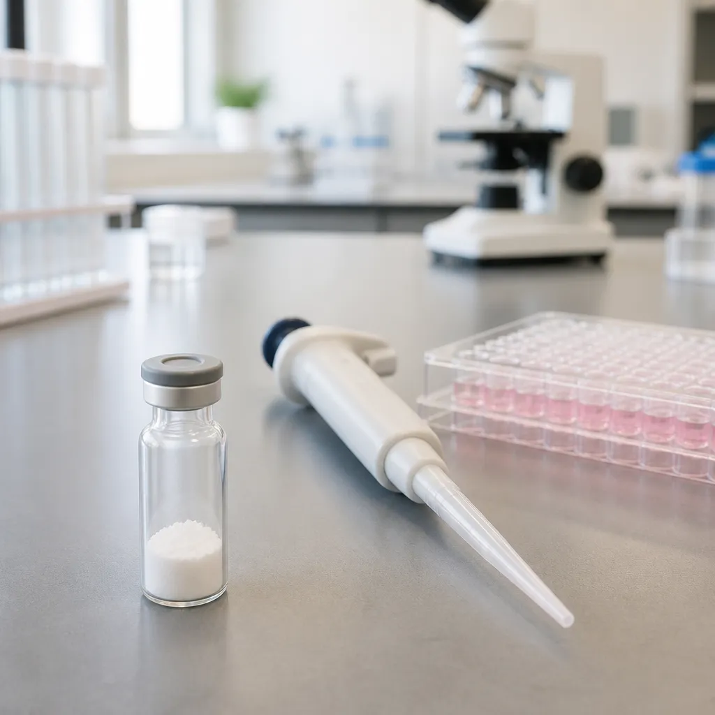
Peptides are not small molecules with negligible protein interaction. Many peptide drugs are engineered to bind albumin or other carriers so they persist longer in circulation, and retatrutide is built on that principle. When the bound fraction is large, the gap between nominal and free concentration can reach several-fold, which is enough to shift an apparent EC50 and make a dose-response curve look like a different compound.
Total versus free concentration
The working equation is straightforward: free concentration equals total concentration multiplied by fu, assuming equilibrium and no other losses. In a well, the total you added is split between a bound pool and a free pool. Serum-free medium may show a fu close to one, while medium containing 10 percent serum can push it substantially lower. The first question I ask on any new retatrutide project is which medium the cells will actually see during the readout.
Why peptides bind plasma proteins
Binding is driven by hydrophobic surfaces, charged residues, and chemical modifications that promote contact with albumin. Retatrutide's reported design includes a modification that promotes reversible noncovalent binding to serum albumin. Reversible binding is usually fast, which means the free fraction is a condition-dependent value rather than a fixed property of the stock vial.
Pro tip: Record two numbers for every retatrutide condition, the nominal total concentration and the calculated free concentration, and state in your methods which one the dose-response axis uses.
What the Published Data Says About Albumin Binding
The most useful public source I have found is a human study on the detection and excretion profile of retatrutide. Its authors describe the molecular modification as promoting reversible noncovalent binding to serum albumin, and they report an extended half-life of roughly six days. Read that half-life as a pharmacokinetic result from a clinical setting, not as an in vitro binding constant you can drop into a spreadsheet.
What albumin binding does and does not tell you
Albumin binding explains why the molecule persists in circulation, but it does not give you a percent bound value for your specific culture medium. Albumin content differs between human plasma, animal serum, and serum-free supplements, and the total protein load in your well depends on the serum percentage you chose. Use the published mechanism to decide whether protein binding is likely to matter, then measure the effect in your own system.
Where the public evidence stops
Public literature on retatrutide is expanding, but protein binding values for research-grade material are still scarce. Most papers focus on receptor pharmacology, weight outcomes, or clinical pharmacokinetics. A review in the open literature summarizes mouse data showing weight reduction and improved glycemic control, which tells you about in vivo activity but nothing about how the peptide partitions in a culture well.
Binding data from a clinical setting describes the body's protein environment, not your well plate. Treat a published binding observation as a hypothesis to test, never as a parameter to copy.
A practical consequence is that a lab should not borrow a fu value from a pharmacokinetic paper and apply it to a 96-well assay without checking the matrix. The mechanism is a reason to test, and the test has to run in the medium your cells will see.
Estimating the Free Fraction Experimentally
Measuring free fraction means separating unbound peptide from protein-bound peptide, then quantifying each pool with an analytical method you trust. Two approaches dominate. Ultrafiltration uses a membrane with a molecular weight cutoff well below albumin's roughly 66 kDa mass, so the protein is retained and the filtrate contains free peptide. Equilibrium dialysis separates the two pools across a semipermeable membrane and runs longer, but it is less sensitive to device artifacts when it is set up correctly.
Ultrafiltration and equilibrium dialysis in practice
Spike retatrutide into the serum-containing matrix at your working concentration, incubate to equilibrium, then spin or dialyze the sample. Quantify the filtrate or dialysate by a validated LC-MS or immunoassay method, and compare it with an unspun control to get recovery. Ultrafiltration is fast and cheap, but the result is only as good as your recovery data. Equilibrium dialysis takes longer, and you must show that equilibrium was reached, usually by sampling at two or more time points.
Using a fixed-ligand binding design as a template
An FDA clinical pharmacology review from a 2022 application file, which is not a retatrutide-specific document, illustrates a useful design. It held a fluorescent ligand at a fixed concentration and measured binding across a range of protein concentrations. You can adapt that logic by keeping the peptide concentration constant, varying serum or albumin across a series, and fitting a saturable binding curve. This design reveals whether binding saturates, which matters because a linear assumption can badly misestimate free concentration at high protein loads.
Pro tip: Pre-condition ultrafiltration membranes with blank matrix, and run a no-protein control through the same device, so you can separate peptide lost to the membrane from peptide that is truly bound to protein.
Serum-Containing Media and Effective Concentration
Once you have a measured fu, the calculation becomes the easy part. The harder part is deciding which medium to use for each experiment and making sure the free fraction you measured is the one the cells experience during the entire incubation. Serum is a moving target, and many assays run for days rather than minutes.
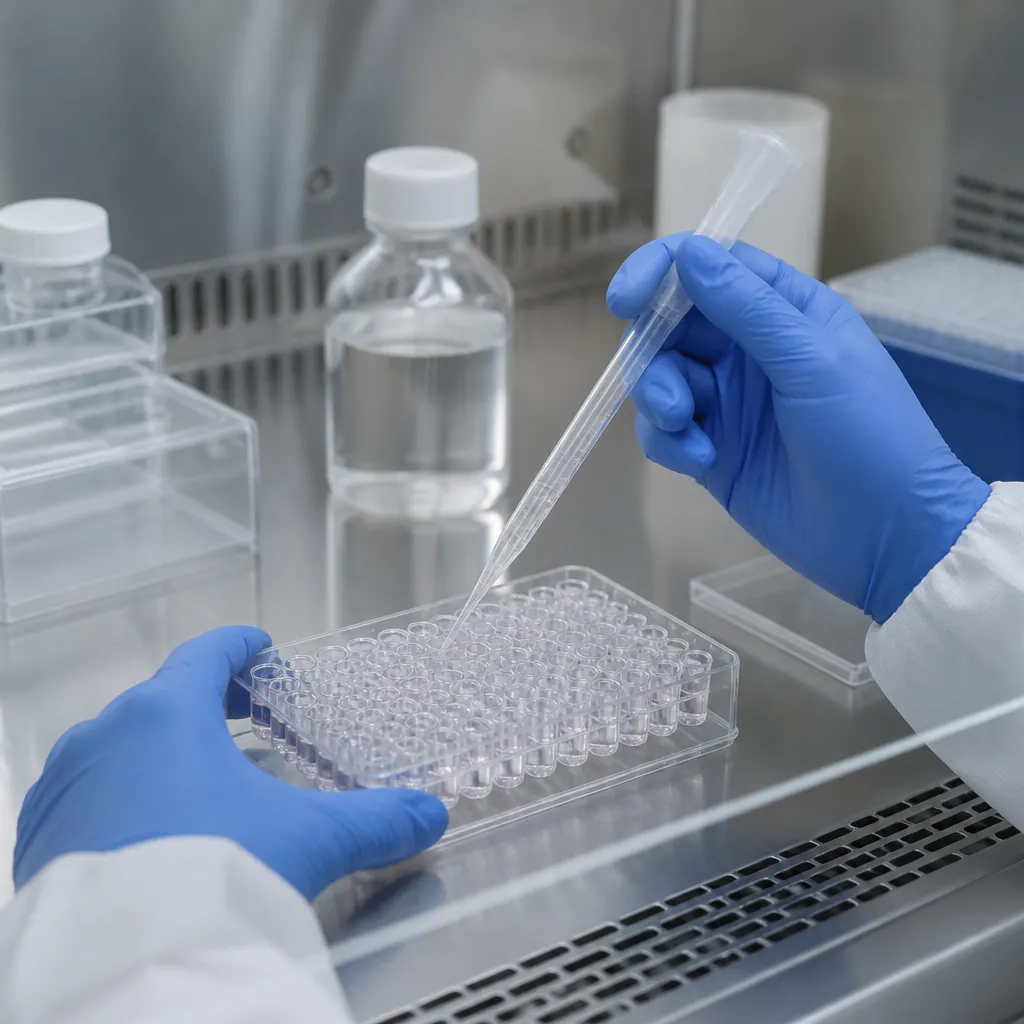
Calculating effective concentration in practice
Use the relationship effective concentration equals total concentration multiplied by fu. Suppose you added 10 nM retatrutide to a medium where your measured fu is 0.2, a hypothetical value used here only for illustration. The free concentration is then 2 nM, and that is the value your dose-response analysis should report. If you plot against 10 nM, every EC50 you calculate will be off by a factor of five, and the error will look like biology.
Serum lot variation and time-dependent shifts
Serum lots differ in protein composition and concentration, so two bottles labeled the same can produce different free fractions. Record the lot number for every experiment, and for binding-sensitive readouts, either test fu per lot or reduce serum where your cell line tolerates it. Cells can also alter the protein environment over time by secreting proteins or consuming components, so a fu measured at time zero may not hold at 72 hours. Where possible, sample the medium at the end of the assay as well as at the start.
Keep in mind that a shift in apparent potency does not automatically mean a change in receptor biology. Before you publish a rightward shift, confirm that protein binding explains it by rerunning the same curve at a lower serum percentage.
Common Calculation Errors in Retatrutide Cell Culture Work
The most common error is treating nominal concentration as free concentration across every condition. The second most common is assuming that a single fu value applies to a whole study, regardless of serum content, incubation time, or plate format. Both errors are invisible in the raw data, which is why they persist through peer review and internal lab meetings.
Assuming nominal concentration equals free concentration
This shortcut works only when protein binding is negligible in your matrix, and you should prove that rather than assume it. A quick test is to compare the potency of the same compound in serum-free and serum-containing medium. If the curves overlay, binding is probably not limiting. If the serum-containing curve shifts to the right, the free fraction is a candidate explanation, and it deserves a direct measurement.
Adsorption to labware and reconstitution choices
Peptides can stick to polypropylene, polystyrene, and filter membranes, which removes material before the cells see it. Use low-binding plastics for stock and intermediate dilutions, and check recovery from your working vessel. Reconstitution also matters. Bacteriostatic water contains benzyl alcohol, so calculate its final concentration in each well and include a vehicle control that matches it. A diluent effect can masquerade as a binding effect if you do not separate the two.
Pro tip: Prepare intermediate dilutions in the same matrix as the final assay wherever feasible, because a stock diluted into buffer and then spiked into serum can behave differently from a stock prepared directly in the working medium.
Documenting Inputs With COA and Batch Records
A binding estimate is only as useful as the material behind it. Lyophilized peptide weighed on a balance contains more than the active peptide, because counterions and residual water contribute to the mass. The certificate of analysis gives you the purity and the information you need to convert a weighed amount into an active concentration. Pepura Labs supplies research-grade retatrutide with a certificate of analysis, Chain of Custody documentation, and batch traceability, and those records let you tie each binding result to a specific lot. You can review the available materials at pepuralabs.ca.

Linking purity and net peptide content to your data
Write the batch number, the stated purity, and the net peptide content into your protocol before the first experiment. If your estimate of active concentration depends on net content, state the correction in the methods, so a reviewer or a colleague can repeat your arithmetic. Canadian laboratory testing can add an independent check on identity and purity when a project depends on tight numbers.
Building a lot-level binding record
Keep a simple table for each lot with the measured fu, the matrix, the serum percentage, the incubation time, and the analytical method. Over several experiments this becomes a practical reference that shows whether binding varies between lots, between serum bottles, or between plate formats. It also protects you when a new batch behaves differently, because you can compare the new fu against the old one rather than guessing.
Pro tip: When a new lot shows a different dose-response curve, repeat the fu measurement in the same serum bottle before concluding that the peptide itself has changed.
Comparing Free Fraction Estimation Approaches
No single method is correct for every lab. The right choice depends on how quickly you need an answer, how much material you can spare, and how precise your downstream dose-response analysis must be. The table below compares three approaches I see used most often in practice.
| Approach | Strength | Main Limitation |
|---|---|---|
| Ultrafiltration with recovery controls | Fast, low equipment demand, easy to run in batches | Membrane adsorption and dilution artifacts can distort fu if no-protein controls are skipped |
| Equilibrium dialysis | Closer to true equilibrium with less device artifact when set up correctly | Slow, requires proof of equilibrium at multiple time points, and uses more material |
| Literature-based assumed fu | Immediate, requires no bench work | Values from clinical or other matrices may not match your medium, so it is only a starting hypothesis |
For most cell culture projects, I recommend ultrafiltration as the first pass, validated with recovery and no-protein controls. Equilibrium dialysis earns its extra time when the binding estimate will anchor a publication or a regulatory-style package. Using an assumed fu from the literature is acceptable only as a planning number, and it should be replaced with a measured value before any quantitative conclusion is drawn.
Whatever method you choose, report it in full. State the matrix, the serum percentage, the time of sampling, the analytical method, and how you corrected for recovery. A free fraction without those details is a number without a context, and in cell culture the context determines whether the result is useful at all.
Frequently Asked Questions
Why does serum protein binding matter for retatrutide in cell culture?
Only the unbound fraction is generally available to interact with receptors in a simple equilibrium model. If a meaningful share of retatrutide binds serum proteins, the effective concentration in your well will be lower than the nominal value you added, and your EC50 estimates will be shifted accordingly.
Can I use the free fraction from a clinical paper for my in vitro assay?
Treat it as a hypothesis, not a parameter. Clinical binding and pharmacokinetic data reflect the human protein environment, while your assay depends on the serum percentage, medium composition, and incubation time you chose. Measure fu in the matrix you actually use.
Which method should a lab start with for measuring free fraction?
Ultrafiltration is a practical first choice because it is fast and runs in batches. Always include recovery controls and a no-protein control through the same device, so you can separate true protein binding from membrane adsorption.
How much does serum lot variation affect the result?
It can matter a great deal, because serum lots differ in protein content and composition. Record lot numbers, test fu for binding-sensitive experiments, and consider reducing serum where your cell line tolerates it.
Does the certificate of analysis change how I calculate effective concentration?
Yes. The certificate of analysis provides purity and related information that you need to convert a weighed amount of lyophilized peptide into an active concentration. Record the batch number and the correction in your methods so the calculation can be reproduced.
What free fraction results have you measured for peptides in your own serum-containing media, and which method gave you the most reproducible numbers? Share your approach or results in the comments so other researchers can compare notes.
References
- Review of retatrutide efficacy and safety data across preclinical and clinical studies
- Human study on retatrutide detection, albumin binding, and excretion profile
- FDA clinical pharmacology review illustrating a fixed-ligand protein binding assay design
- Review of GLP-1 receptor mechanisms and signaling pathways