Many metabolic peptide protocols track glucose, lipid or body composition endpoints and add oxidative stress readouts as an afterthought. That gap matters. Reactive oxygen species (ROS) sit upstream of many of the cellular changes researchers are trying to explain, so an oxidative stress assay peptide study that skips them can misread a metabolic effect. This article explains how to add ROS measurement to in vitro retatrutide protocols, which readouts deserve trust, which need caution, and how to document the compound so your oxidative data can be reproduced.
Table of Contents
- Quick Takeaways
- Why Oxidative Endpoints Matter in Metabolic Studies
- Choosing a ROS Readout That Holds Up
- Oxidative Damage Markers as Complementary Endpoints
- Antioxidant Enzyme Readouts: SOD, Catalase and GPx
- Designing a Retatrutide Cell Culture Protocol With ROS Endpoints
- Comparing ROS Measurement Approaches Side by Side
- Interpreting Results and Documenting the Compound
- Frequently Asked Questions
- References
Quick Takeaways
| Key Insight | Explanation |
|---|---|
| Measure at least two readouts | A single ROS dye cannot separate oxidant production from probe artifacts. Pair a ROS signal with an oxidative damage marker or an antioxidant enzyme panel. |
| Run cell-free background wells | Dyes can react with medium components and with the compound itself. Include a no-cell well containing compound, probe and medium, and subtract it before interpreting treatment effects. |
| Match the endpoint to the timeline | ROS signals can change within minutes, while lipid damage builds over hours. One timepoint rarely answers the question. |
| Normalize to viability and protein | Oxidative signal per well shifts when cell number changes. Normalize to a viability readout or total protein from the same well. |
| Record the lot, not just the vial | Lot numbers and certificate of analysis references let you separate a compound effect from a lot-to-lot difference. |
| Report probe, settings and blanks | Without the probe identity, instrument settings and blank values, an oxidative result cannot be compared across labs or repeated by a collaborator. |
Why Oxidative Endpoints Matter in Metabolic Studies
Metabolic peptides change how cells handle fuel, and fuel handling runs through mitochondria. When electron transport leaks, superoxide and other reactive species form, so redox readouts often move alongside glucose and lipid measurements. A study that records only the metabolic endpoints misses that link.
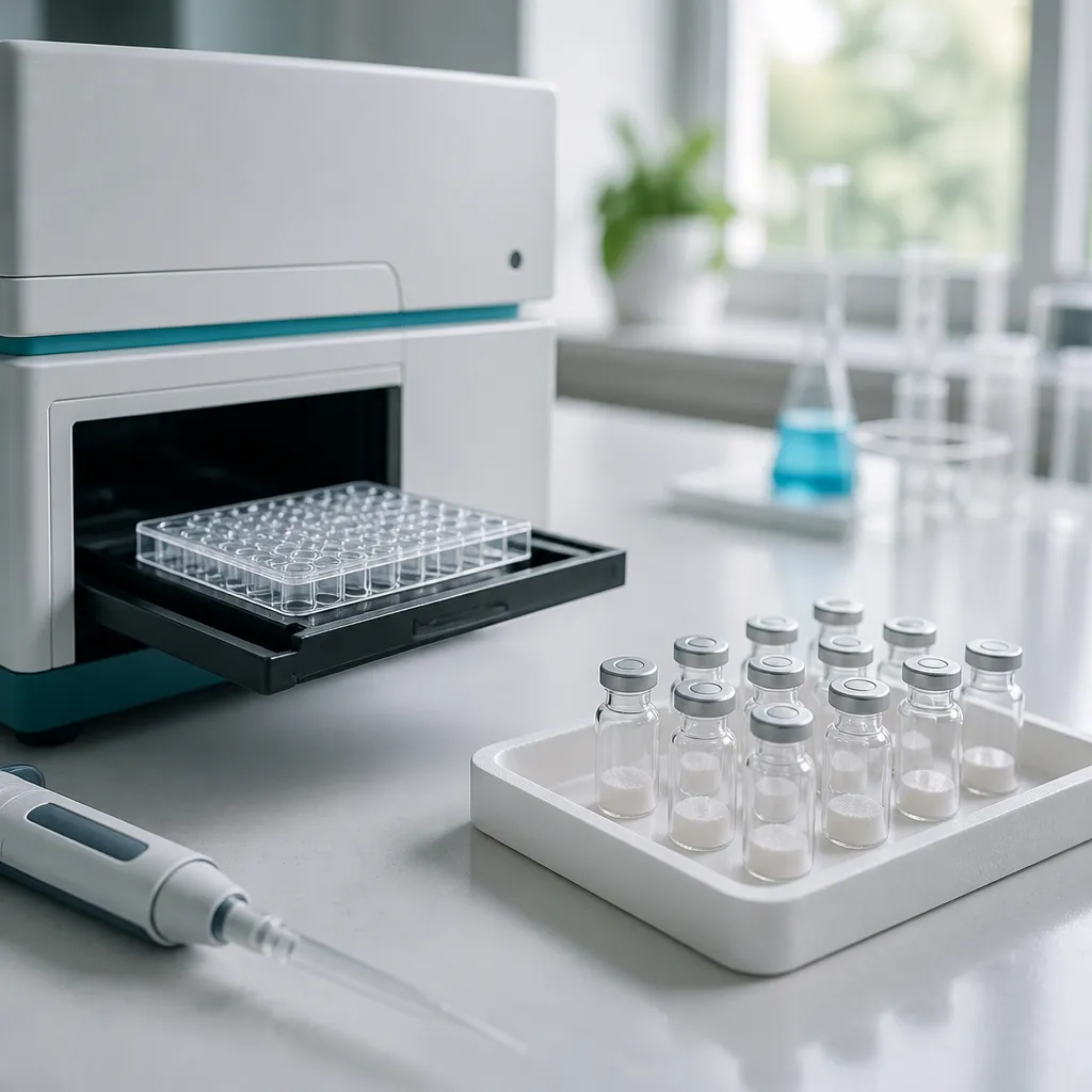
In practice, the most common design problem is that oxidative endpoints get added after the metabolic data are already collected. That order makes it hard to say whether a ROS change drives the metabolic effect, follows it, or is simply assay noise. Plan the redox readout at the protocol stage, with its own controls, timepoints and sample handling.
Setting the Question Before the Assay
Because a multi-target peptide can shift several pathways at once, a single ROS number rarely explains the whole picture. Write down which pathway you expect to change before you pick a readout. A hypothesis about mitochondrial leak points to different probes and timepoints than a hypothesis about antioxidant enzyme induction.
The 2022 guidelines paper by M. P. Murphy and colleagues on measuring reactive oxygen species and oxidative damage in cells and in vivo is a useful starting point because it treats those as separate measurements. Your protocol should do the same. A ROS signal and a damage marker answer different questions, and presenting one as evidence for the other weakens a paper quickly.
Choosing a ROS Readout That Holds Up
Choosing a ROS readout is the decision that most determines whether your data survive review. Each method reports something specific, and none reports "ROS" in a general sense. Pick the readout that matches the species and compartment you care about.
Fluorescent Probes and Their Artifacts
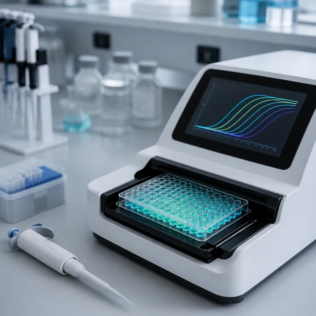
Chemical fluorescent probes are the default in many labs because they are inexpensive, fast and compatible with plate readers. The catch is that many of them are oxidized by more than one species and can gain signal from light exposure or spontaneous oxidation in the medium. A rising fluorescence trace in a well with no cells is a warning sign, not a result.
A fluorescence increase tells you that something oxidized the probe. It does not tell you which oxidant did it, or where it happened.
Run a cell-free well with the compound, the probe and the medium, then subtract that background before interpreting any treatment effect. If the cell-free signal moves with concentration, the assay is measuring chemistry rather than biology. That single control has saved more datasets in our experience than any statistical correction.
Genetically Encoded Redox Sensors
Genetically encoded sensors report redox state inside defined compartments, which chemical dyes cannot do. They require transfection or a stable cell line, and expression level can change baseline readings. Build in a validation step that confirms sensor expression is comparable across treatment groups.
The trade-off is time. Sensor lines take weeks to establish, so most teams use them for mechanistic follow-up after a dye screen has produced a signal worth chasing.
Oxidative Damage Markers as Complementary Endpoints
Damage markers answer a different question. Instead of asking whether oxidants are present now, they ask whether lipids, proteins or DNA have been modified by oxidation over the course of the experiment. That makes them slower to produce, but more durable and less sensitive to a single transient spike.
Lipid Peroxidation Readouts
Lipid peroxidation products such as malondialdehyde and 4-hydroxynonenal accumulate after oxidant exposure, which is why they are widely measured. Thiobarbituric acid reactive substance assays are known to react with several aldehydes, so their specificity is limited. Validate any kit against a known oxidant positive control before running your full treatment matrix.
Lipid peroxidation also builds over hours. A 30-minute read will miss it entirely, so schedule these measurements at the later timepoints of your design rather than at the end of a short exposure.
Protein Carbonyls and DNA Lesions
Protein carbonyl assays detect oxidized amino acid side chains and are a reasonable choice when you want a cumulative readout from lysates. DNA lesion markers are more labour-intensive and need careful extraction to avoid oxidation during sample prep. In practice, the sample prep step is where many oxidative damage numbers are quietly inflated.
Do not compare damage markers across different harvest times without time-matched vehicle controls. Oxidation can continue in lysates left on the bench, so process every group on the same schedule and record the delay between harvest and freezing.
Antioxidant Enzyme Readouts: SOD, Catalase and GPx
Antioxidant enzyme activity tells you about the defense side of the balance. If a compound shifts ROS and enzyme activity in the same direction, the interpretation is more coherent than either measurement alone. Most reviews of oxidative stress frame the problem as an imbalance between ROS production and endogenous defenses, and your protocol should test that framing directly.

A 2021 review by Q. Qiao and colleagues on dietary bioactive peptides and redox balance describes peptides that reduced oxidative stress by enhancing superoxide dismutase, catalase and glutathione peroxidase. Those findings concern food-derived peptides rather than retatrutide, so treat them as hypotheses for your enzyme panel, not as evidence for the compound you are testing.
Superoxide Dismutase and Catalase
Superoxide dismutase and catalase assays are sensitive to sample handling. Lysis buffer composition, protease inhibitors and time on ice all change apparent activity, and so does the protein normalization method. Run these assays on fresh lysates from the same batch, and keep buffer composition identical across groups.
Glutathione Peroxidase and Reduced Glutathione
Glutathione peroxidase activity is usually coupled to a secondary enzyme reaction, which makes it vulnerable to any compound that interferes with the coupling step. Reduced to oxidized glutathione ratios are a useful complement, but they are sensitive to thiol-containing reagents in your buffers. Check for interference with a spike-in standard before you trust a ratio.
A 2022 study by N. Rodríguez-Arana and colleagues in an MDPI journal reports that the radical scavenging properties of certain peptides could account for protective effects against oxidative damage in its experimental system. That is a mechanistic idea worth testing with your own data, not a result you can transfer to a metabolic peptide without it.
Designing a Retatrutide Cell Culture Protocol With ROS Endpoints
Start with the question, then build the matrix. A useful design compares vehicle, a concentration series of retatrutide, and a positive oxidant control at a dose your cell model tolerates. Without the positive control, a null result tells you nothing about whether the assay could have detected a change at all.
Controls That Separate Compound Effects From Assay Effects
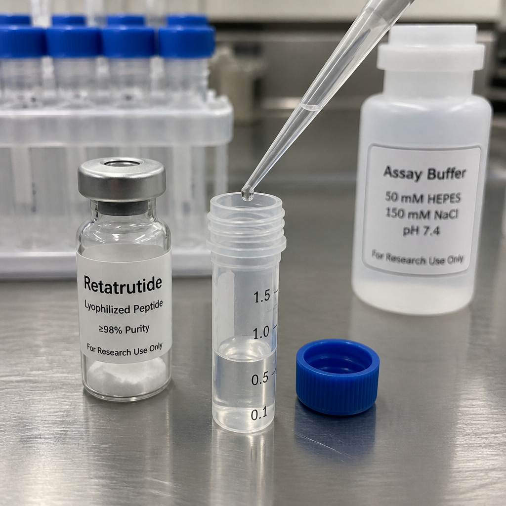
Your vehicle control must match the diluent exactly. Bacteriostatic water contains a preservative, so if you reconstitute lyophilized retatrutide with it, the vehicle well needs the same bacteriostatic water at the same final concentration. Otherwise you are comparing the compound against plain medium and attributing the preservative's effect to the peptide.
Pro tip: Make a single bulk vehicle stock and dilute it into every vehicle well, including the cell-free background wells. Any variation in the vehicle then cancels out of your treatment comparisons instead of hiding inside them.
Timing, Sample Handling and Light Exposure
Pick at least three timepoints that span early and late responses, because transient ROS spikes and slower damage accumulation rarely line up on a single read. Protect fluorescent probes and plates from light between loading and reading. Process all groups in parallel, and record the minutes between harvest and freezing for every well.
Reconstitute once, aliquot into single-use volumes, and avoid repeated freeze-thaw cycles. Each cycle is a variable you cannot easily reconstruct later, so write the thaw count directly on the tube label.
Comparing ROS Measurement Approaches Side by Side
The table below summarizes the three approaches we see most often in metabolic peptide protocols. None is the right answer on its own. The strongest designs usually combine one fast, inexpensive readout with one slower, more specific readout.
| Approach | Best Use | Main Limitation |
|---|---|---|
| Chemical fluorescent probes | Fast plate-based screening and dose-response work | Low specificity, sensitive to light and cell-free chemistry |
| Genetically encoded redox sensors | Compartment-specific mechanistic follow-up | Requires transfection or stable lines, and expression can shift the baseline |
| Oxidative damage marker panels (lipid peroxidation, protein carbonyls) | Cumulative damage over hours to days | Slower, prone to sample-prep artifacts, and kit specificity varies |
If you can afford only one readout, use a chemical probe for screening and add a damage marker for the figure that goes into a manuscript. Reviewers tend to trust a fast signal that is backed by an independent, slower endpoint more than a single dye curve.
Enzyme activity panels sit between these categories. They add a defense-side readout that helps interpret the other two, especially when a probe signal rises but damage markers do not follow within your observation window.
Interpreting Results and Documenting the Compound
Interpretation starts with normalization. Oxidative signal per well changes when cell number changes, so divide by a viability readout or total protein measured from the same well. Report both raw and normalized values, because collaborators will want to check your normalization choice.

Normalizing Oxidative Signal
Normalize within plate and within batch first, then compare batches. A dose response that looks clear on one plate can collapse when plates are pooled, because edge effects and temperature gradients vary. Include the same reference well on every plate so you can detect drift.
Documenting the Compound Lot
Oxidative data are only as reproducible as the compound behind them. Pepura Labs supplies lyophilized retatrutide with Canadian laboratory verification and Chain of Custody documentation, and each batch is traceable to its certificate of analysis. Record the lot number, the COA reference, the reconstitution date and the storage history alongside every oxidative dataset.
Pro tip: Keep the COA for each lot in the same folder as the raw plate reader exports. When a result shifts between runs, you can check the lot first instead of reconstructing the chain of custody from memory.
Storage matters more than shipping, but arrival records still belong in your file. Canadian research teams ordering from us receive Xpresspost delivery, so log the arrival date and the storage conditions your team used on receipt.
Frequently Asked Questions
Can I run an oxidative stress assay peptide study with a single ROS dye?
You can, but a single dye gives weak evidence on its own. Pair it with a cell-free background well and at least one damage marker or enzyme readout so the result does not depend on one chemistry.
How many timepoints should a ROS protocol include?
Use at least three timepoints that span early and late responses. ROS signals and lipid damage rarely peak at the same time, so a single read can make a real effect look absent.
Which control matters most in a retatrutide ROS experiment?
The vehicle control matched to your reconstitution diluent matters most, followed by the cell-free background well. If your diluent contains a preservative, the vehicle well must contain it too.
Does a ROS result in cell culture predict an effect in people?
No. Pepura Labs supplies retatrutide for in vitro research only. Cell-based oxidative results describe a model system and do not translate directly to human outcomes.
What documentation should accompany an oxidative dataset?
Record the lot number, the COA reference, the reconstitution date, the number of freeze-thaw cycles, the probe identity, instrument settings and blank values. Without these, a collaborator cannot repeat the run.
Should I normalize to viability or to protein?
Use whichever readout you can measure from the same well, and state the choice in your methods. If you normalize to viability, confirm that treatment does not change viability enough to distort the oxidative signal.
If you have run ROS or oxidative damage assays on metabolic peptides, share which probe, controls and timepoints gave you the most reproducible results and where your protocol still breaks down.
We would love your feedback and any insights you would share with others. What perspective would you add?
References
- Guidelines for measuring reactive oxygen species and oxidative damage in cells and in vivo
- Review of dietary bioactive peptides and their role in redox balance
- Study on peptide protection against oxidative stress and metabolic damage
- Overview of food-derived bioactive peptides as functional ingredients